 Movie
Movie Controller

[English] 日本語
 Yorodumi
- PDB-6cbl: x-ray structure of NeoB from Streptomyces fradiae in complex with... -
+ Open data

- Basic information
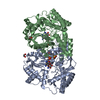
| Entry | Database: PDB / ID: 6cbl | ||||||
|---|---|---|---|---|---|---|---|
| Title | x-ray structure of NeoB from Streptomyces fradiae in complex with neamine as an external aldimine | ||||||
 Components Components | Neamine transaminase NeoN | ||||||
 Keywords Keywords | TRANSFERASE / Neomycin / aminotransferase | ||||||
| Function / homology |  Function and homology information Function and homology informationneamine transaminase / neomycin C transaminase / neomycin biosynthetic process / transaminase activity / pyridoxal phosphate binding Similarity search - Function | ||||||
| Biological species |  Streptomyces fradiae (bacteria) Streptomyces fradiae (bacteria) | ||||||
| Method |  X-RAY DIFFRACTION / SYNCHROTRON / MOLECULAR REPLACEMENT / Resolution: 1.6 Å X-RAY DIFFRACTION / SYNCHROTRON / MOLECULAR REPLACEMENT / Resolution: 1.6 Å | ||||||
 Authors Authors | Thoden, J.B. / Dow, G.T. / Holden, H.M. | ||||||
| Funding support |  United States, 1items United States, 1items
| ||||||
 Citation Citation | Journal: Protein Sci. / Year: 2018 Title: The three-dimensional structure of NeoB: An aminotransferase involved in the biosynthesis of neomycin. Authors: Dow, G.T. / Thoden, J.B. / Holden, H.M. | ||||||
| History |
|
- Structure visualization
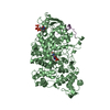
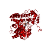
| Structure viewer | Molecule: MolmilJmol/JSmol |
|---|
- Downloads & links
-Download
| PDBx/mmCIF format | 6cbl.cif.gz | 670.1 KB | Display | PDBx/mmCIF format |
|---|---|---|---|---|
| PDB format | pdb6cbl.ent.gz | 550.5 KB | Display | PDB format |
| PDBx/mmJSON format | 6cbl.json.gz | Tree view | PDBx/mmJSON format | |
| Others |  Other downloads Other downloads |
-Validation report
| Arichive directory | https://data.pdbj.org/pub/pdb/validation_reports/cb/6cblftp://data.pdbj.org/pub/pdb/validation_reports/cb/6cbl | HTTPS FTP |
|---|
-Related structure data
| Related structure data |  6cbkSC  6cbmC  6cbnC  6cboC S: Starting model for refinement C: citing same article ( |
|---|---|
| Similar structure data |


-Links
 PDBj
PDBj- Assembly
| Deposited unit | 
| ||||||||
|---|---|---|---|---|---|---|---|---|---|
| 1 | 
| ||||||||
| 2 | 
| ||||||||
| 3 | 
| ||||||||
| 4 | 
| ||||||||
| Unit cell |
|
-Components
| #1: Protein | Mass: 45766.520 Da / Num. of mol.: 8 Source method: isolated from a genetically manipulated source Source: (gene. exp.) Streptomyces fradiae (bacteria) / Gene: neoN, neo18, neoB / Production host: References: UniProt: Q53U08, neamine transaminase, neomycin C transaminase #2: Chemical | ChemComp-DOW / (   Mass: 553.501 Da / Num. of mol.: 8 / Source method: obtained synthetically / Formula: C20H36N5O11P Mass: 553.501 Da / Num. of mol.: 8 / Source method: obtained synthetically / Formula: C20H36N5O11P#3: Chemical | ChemComp-CL / |   Mass: 35.453 Da / Num. of mol.: 1 / Source method: obtained synthetically / Formula: Cl Mass: 35.453 Da / Num. of mol.: 1 / Source method: obtained synthetically / Formula: Cl#4: Water | ChemComp-HOH / |  Mass: 18.015 Da / Num. of mol.: 2470 / Source method: isolated from a natural source / Formula: H2O Mass: 18.015 Da / Num. of mol.: 2470 / Source method: isolated from a natural source / Formula: H2O |
|---|
-Experimental details
-Experiment
| Experiment | Method: X-RAY DIFFRACTION / Number of used crystals: 1 |
|---|
- Sample preparation
| Crystal | Density Matthews: 2.22 Å3/Da / Density % sol: 44.62 % |
|---|---|
| Crystal grow | Temperature: 293 K / Method: vapor diffusion, hanging drop / pH: 7.5 Details: 16-21% PEG-3350, 300 mM KCl, 100 mM HEPES, 5 mM neamine, 1 mM PLP |
-Data collection
| Diffraction | Mean temperature: 100 K |
|---|---|
| Diffraction source | Source: SYNCHROTRON / Site: APS / Beamline: 19-BM / Wavelength: 0.9498 Å |
| Detector | Type: ADSC QUANTUM 210r / Detector: CCD / Date: Jun 10, 2017 |
| Radiation | Protocol: SINGLE WAVELENGTH / Monochromatic (M) / Laue (L): M / Scattering type: x-ray |
| Radiation wavelength | Wavelength: 0.9498 Å / Relative weight: 1 |
| Reflection | Resolution: 1.6→50 Å / Num. obs: 367186 / % possible obs: 87.2 % / Observed criterion σ(F): 0 / Observed criterion σ(I): 0 / Redundancy: 3.9 % / Rmerge(I) obs: 0.049 / Rsym value: 0.049 / Net I/σ(I): 38.4 |
| Reflection shell | Resolution: 1.6→1.66 Å / Redundancy: 2.6 % / Rmerge(I) obs: 0.1 / Mean I/σ(I) obs: 7.9 / Num. unique obs: 29397 / Rsym value: 0.1 / % possible all: 70 |
- Processing
| Software |
| ||||||||||||||||||||||||||||||||||||||||||||||||||||||||||||||||||||||||||||||||||||||||||||||||||||||||||||||||||||||||||||||||||||||||||||||||||||||||||||||||||||||||||||||||||||||
|---|---|---|---|---|---|---|---|---|---|---|---|---|---|---|---|---|---|---|---|---|---|---|---|---|---|---|---|---|---|---|---|---|---|---|---|---|---|---|---|---|---|---|---|---|---|---|---|---|---|---|---|---|---|---|---|---|---|---|---|---|---|---|---|---|---|---|---|---|---|---|---|---|---|---|---|---|---|---|---|---|---|---|---|---|---|---|---|---|---|---|---|---|---|---|---|---|---|---|---|---|---|---|---|---|---|---|---|---|---|---|---|---|---|---|---|---|---|---|---|---|---|---|---|---|---|---|---|---|---|---|---|---|---|---|---|---|---|---|---|---|---|---|---|---|---|---|---|---|---|---|---|---|---|---|---|---|---|---|---|---|---|---|---|---|---|---|---|---|---|---|---|---|---|---|---|---|---|---|---|---|---|---|---|
| Refinement | Method to determine structure: MOLECULAR REPLACEMENT Starting model: 6cbk Resolution: 1.6→30 Å / Cor.coef. Fo:Fc: 0.947 / Cor.coef. Fo:Fc free: 0.919 / SU B: 2.397 / SU ML: 0.081 / Cross valid method: THROUGHOUT / ESU R: 0.114 / ESU R Free: 0.117 / Details: HYDROGENS HAVE BEEN ADDED IN THE RIDING POSITIONS
| ||||||||||||||||||||||||||||||||||||||||||||||||||||||||||||||||||||||||||||||||||||||||||||||||||||||||||||||||||||||||||||||||||||||||||||||||||||||||||||||||||||||||||||||||||||||
| Solvent computation | Ion probe radii: 0.8 Å / Shrinkage radii: 0.8 Å / VDW probe radii: 1.2 Å | ||||||||||||||||||||||||||||||||||||||||||||||||||||||||||||||||||||||||||||||||||||||||||||||||||||||||||||||||||||||||||||||||||||||||||||||||||||||||||||||||||||||||||||||||||||||
| Displacement parameters | Biso mean: 23.734 Å2
| ||||||||||||||||||||||||||||||||||||||||||||||||||||||||||||||||||||||||||||||||||||||||||||||||||||||||||||||||||||||||||||||||||||||||||||||||||||||||||||||||||||||||||||||||||||||
| Refinement step | Cycle: 1 / Resolution: 1.6→30 Å
| ||||||||||||||||||||||||||||||||||||||||||||||||||||||||||||||||||||||||||||||||||||||||||||||||||||||||||||||||||||||||||||||||||||||||||||||||||||||||||||||||||||||||||||||||||||||
| Refine LS restraints |
|