Resolution: 1.6→1.66 Å / Redundancy: 1.9 % / Rmerge(I) obs: 0.111 / Mean I/σ(I) obs: 10.5 / Num. unique obs: 18111 / Rsym value: 0.111 / % possible all: 83.4
-
Processing
Software
Name
Version
Classification
REFMAC
5.8.0124
refinement
HKL-3000
datareduction
HKL-3000
datascaling
PHASER
phasing
Refinement
Method to determine structure: SAD / Resolution: 1.6→50 Å / Cor.coef. Fo:Fc: 0.959 / Cor.coef. Fo:Fc free: 0.944 / SU B: 1.42 / SU ML: 0.05 / Cross valid method: THROUGHOUT / ESU R: 0.085 / ESU R Free: 0.086 / Details: HYDROGENS HAVE BEEN ADDED IN THE RIDING POSITIONS
Rfactor
Num. reflection
% reflection
Selection details
Rfree
0.18818
10040
5 %
RANDOM
Rwork
0.15521
-
-
-
obs
0.15685
191197
92.16 %
-
Solvent computation
Ion probe radii: 0.8 Å / Shrinkage radii: 0.8 Å / VDW probe radii: 1.2 Å
 Movie
Movie Controller

 Yorodumi
 Open data
 Basic information
 Components
 Keywords
 Function and homology information
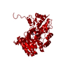
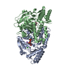
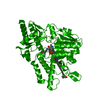
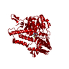
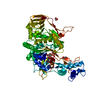
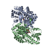
 Micromonospora echinospora (bacteria)
 X-RAY DIFFRACTION /
  Authors
 United States, 1items
  Citation
 Structure visualization
 Downloads & links
 Other downloads


 PDBj
PDBj Assembly
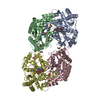
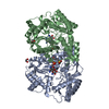


 Mass: 209.263 Da / Num. of mol.: 3 / Source method: obtained synthetically / Formula: C7H15NO4S / Comment: pH buffer*YM
 Mass: 62.068 Da / Num. of mol.: 4 / Source method: obtained synthetically / Formula: C2H6O2
 Mass: 553.501 Da / Num. of mol.: 4 / Source method: obtained synthetically / Formula: C20H36N5O11P
 Mass: 96.063 Da / Num. of mol.: 2 / Source method: obtained synthetically / Formula: SO4
 Sample preparation
 Processing