National Institutes of Health/National Institute Of Allergy and Infectious Diseases (NIH/NIAID)
R01-AI103171
United States
Citation
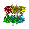
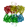
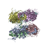
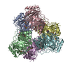
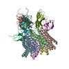
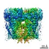
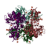
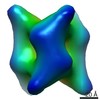
Journal: Nature / Year: 2018 Title: Cryo-EM structure of the insect olfactory receptor Orco. Authors: Joel A Butterwick / Josefina Del Mármol / Kelly H Kim / Martha A Kahlson / Jackson A Rogow / Thomas Walz / Vanessa Ruta / Abstract: The olfactory system must recognize and discriminate amongst an enormous variety of chemicals in the environment. To contend with such diversity, insects have evolved a family of odorant-gated ion ...The olfactory system must recognize and discriminate amongst an enormous variety of chemicals in the environment. To contend with such diversity, insects have evolved a family of odorant-gated ion channels comprised of a highly conserved co-receptor (Orco) and a divergent odorant receptor (OR) that confers chemical specificity. Here, we present the single-particle cryo-electron microscopy structure of an Orco homomer from the parasitic fig wasp Apocrypta bakeri at 3.5 Å resolution, providing structural insight into this receptor family. Orco possesses a novel channel architecture, with four subunits symmetrically arranged around a central pore that diverges into four lateral conduits that open to the cytosol. The Orco tetramer has few inter-subunit interactions within the membrane and is bound together by a small cytoplasmic anchor domain. The minimal sequence conservation among ORs maps largely to the pore and anchor domain, shedding light on how the architecture of this receptor family accommodates its remarkable sequence diversity and facilitates the evolution of odour tuning.
History
Deposition
Jan 19, 2018
Deposition site: RCSB / Processing site: RCSB
Revision 1.0
Aug 22, 2018
Provider: repository / Type: Initial release
Revision 1.0
Aug 22, 2018
Data content type: EM metadata / Data content type: EM metadata / Provider: repository / Type: Initial release
Revision 1.0
Aug 22, 2018
Data content type: Image / Data content type: Image / Provider: repository / Type: Initial release
Revision 1.0
Aug 22, 2018
Data content type: Primary map / Data content type: Primary map / Provider: repository / Type: Initial release
Revision 1.0
Aug 22, 2018
Data content type: EM metadata / Data content type: EM metadata / Provider: repository / Type: Initial release
Revision 1.0
Aug 22, 2018
Data content type: Image / Data content type: Image / Provider: repository / Type: Initial release
Revision 1.0
Aug 22, 2018
Data content type: Primary map / Data content type: Primary map / Provider: repository / Type: Initial release
Data content type: EM metadata / Data content type: EM metadata / EM metadata / Group: Data processing / Experimental summary / Data content type: EM metadata / EM metadata / Category: em_admin / em_software / Data content type: EM metadata / EM metadata / Item: _em_admin.last_update / _em_software.name
Mass: 53513.238 Da / Num. of mol.: 4 Source method: isolated from a genetically manipulated source Source: (gene. exp.) Apocrypta bakeri (insect) / Gene: Or2 / Production host: Homo sapiens (human) / References: UniProt: B0FAQ4
Has protein modification
N
-
Experimental details
-
Experiment
Experiment
Method: ELECTRON MICROSCOPY
EM experiment
Aggregation state: PARTICLE / 3D reconstruction method: single particle reconstruction
-
Sample preparation
Component
Name: Orco homotetramer bound to four Fab molecules in a detergent micelle Type: COMPLEX / Entity ID: all / Source: RECOMBINANT
Molecular weight
Experimental value: NO
Source (natural)
Organism: Apocrypta bakeri (insect)
Source (recombinant)
Organism: Homo sapiens (human)
Buffer solution
pH: 7.5
Specimen
Embedding applied: NO / Shadowing applied: NO / Staining applied: NO / Vitrification applied: YES
Vitrification
Cryogen name: ETHANE
-
Electron microscopy imaging
Experimental equipment
Model: Titan Krios / Image courtesy: FEI Company
Microscopy
Model: FEI TITAN KRIOS
Electron gun
Electron source: FIELD EMISSION GUN / Accelerating voltage: 300 kV / Illumination mode: FLOOD BEAM
Electron lens
Mode: BRIGHT FIELD
Image recording
Average exposure time: 0.2 sec. / Electron dose: 80 e/Å2 / Detector mode: SUPER-RESOLUTION / Film or detector model: GATAN K2 SUMMIT (4k x 4k) / Num. of grids imaged: 1221 / Num. of real images: 53141
Image scans
Movie frames/image: 50 / Used frames/image: 1-50
-
Processing
Software
Name
Version
Classification
NB
PHENIX
1.13_2998:
refinement
PDB_EXTRACT
3.22
dataextraction
EM software
Name: PHENIX / Category: model refinement
CTF correction
Type: PHASE FLIPPING AND AMPLITUDE CORRECTION
3D reconstruction
Resolution: 3.5 Å / Resolution method: FSC 0.143 CUT-OFF / Num. of particles: 53141 / Symmetry type: POINT
In the structure databanks used in Yorodumi, some data are registered as the other names, "COVID-19 virus" and "2019-nCoV". Here are the details of the virus and the list of structure data.
Jan 31, 2019. EMDB accession codes are about to change! (news from PDBe EMDB page)
EMDB accession codes are about to change! (news from PDBe EMDB page)
The allocation of 4 digits for EMDB accession codes will soon come to an end. Whilst these codes will remain in use, new EMDB accession codes will include an additional digit and will expand incrementally as the available range of codes is exhausted. The current 4-digit format prefixed with “EMD-” (i.e. EMD-XXXX) will advance to a 5-digit format (i.e. EMD-XXXXX), and so on. It is currently estimated that the 4-digit codes will be depleted around Spring 2019, at which point the 5-digit format will come into force.
The EM Navigator/Yorodumi systems omit the EMD- prefix.
Related info.:Q: What is EMD? / ID/Accession-code notation in Yorodumi/EM Navigator
Yorodumi is a browser for structure data from EMDB, PDB, SASBDB, etc.
This page is also the successor to EM Navigator detail page, and also detail information page/front-end page for Omokage search.
The word "yorodu" (or yorozu) is an old Japanese word meaning "ten thousand". "mi" (miru) is to see.
Related info.:EMDB / PDB / SASBDB / Comparison of 3 databanks / Yorodumi Search / Aug 31, 2016. New EM Navigator & Yorodumi / Yorodumi Papers / Jmol/JSmol / Function and homology information / Changes in new EM Navigator and Yorodumi
 Movie
Movie Controller


 Open data
 Basic information
 Components
 Keywords
 Function and homology information
 Apocrypta bakeri (insect)
 Authors
 United States, 1items
  Citation
 Structure visualization
 Downloads & links
 Other downloads
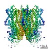
 PDBj
PDBj Assembly
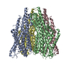
 Homo sapiens (human) / References: UniProt: B0FAQ4
 Sample preparation
 Electron microscopy imaging
 FIELD EMISSION GUN / Accelerating voltage: 300 kV / Illumination mode: FLOOD BEAM
 Processing