 Movie
Movie Controller


+ Open data

- Basic information
| Entry | Database: EMDB / ID: EMD-7352 | |||||||||
|---|---|---|---|---|---|---|---|---|---|---|
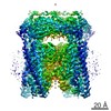
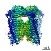
| Title | Cryo-EM structure of Orco | |||||||||
 Map data Map data | Odorant receptor | |||||||||
 Sample Sample |
| |||||||||
 Keywords Keywords | Olfactory receptor / Ion channel / Insect / Fab / MEMBRANE PROTEIN | |||||||||
| Function / homology | Olfactory receptor, insect / 7tm Odorant receptor / olfactory receptor activity / odorant binding / signal transduction / identical protein binding / plasma membrane / Odorant receptor Function and homology information Function and homology information | |||||||||
| Biological species |  Apocrypta bakeri (insect) Apocrypta bakeri (insect) | |||||||||
| Method | single particle reconstruction / cryo EM / Resolution: 3.5 Å | |||||||||
 Authors Authors | Butterwick JA / Kim KH / Walz T / Ruta V | |||||||||
| Funding support |  United States, 1 items United States, 1 items
| |||||||||
 Citation Citation | Journal: Nature / Year: 2018 Title: Cryo-EM structure of the insect olfactory receptor Orco. Authors: Joel A Butterwick / Josefina Del Mármol / Kelly H Kim / Martha A Kahlson / Jackson A Rogow / Thomas Walz / Vanessa Ruta / Abstract: The olfactory system must recognize and discriminate amongst an enormous variety of chemicals in the environment. To contend with such diversity, insects have evolved a family of odorant-gated ion ...The olfactory system must recognize and discriminate amongst an enormous variety of chemicals in the environment. To contend with such diversity, insects have evolved a family of odorant-gated ion channels comprised of a highly conserved co-receptor (Orco) and a divergent odorant receptor (OR) that confers chemical specificity. Here, we present the single-particle cryo-electron microscopy structure of an Orco homomer from the parasitic fig wasp Apocrypta bakeri at 3.5 Å resolution, providing structural insight into this receptor family. Orco possesses a novel channel architecture, with four subunits symmetrically arranged around a central pore that diverges into four lateral conduits that open to the cytosol. The Orco tetramer has few inter-subunit interactions within the membrane and is bound together by a small cytoplasmic anchor domain. The minimal sequence conservation among ORs maps largely to the pore and anchor domain, shedding light on how the architecture of this receptor family accommodates its remarkable sequence diversity and facilitates the evolution of odour tuning. | |||||||||
| History |
|
- Structure visualization
| Movie |
Movie viewer |
|---|---|
| Structure viewer | EM map: SurfViewMolmilJmol/JSmol |
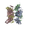
| Supplemental images |

- Downloads & links
-EMDB archive
| Map data | emd_7352.map.gz | 179.4 MB | EMDB map data format | |
|---|---|---|---|---|
| Header (meta data) | emd-7352-v30.xmlemd-7352.xml | 13.9 KB 13.9 KB | Display Display | EMDB header |
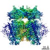
| Images | 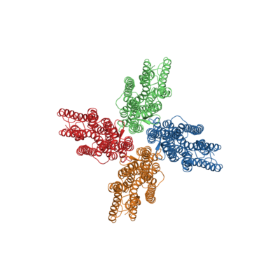 emd_7352.png emd_7352.png | 79.6 KB | ||
| Filedesc metadata | emd-7352.cif.gz | 5.8 KB | ||
| Archive directory |  http://ftp.pdbj.org/pub/emdb/structures/EMD-7352ftp://ftp.pdbj.org/pub/emdb/structures/EMD-7352 http://ftp.pdbj.org/pub/emdb/structures/EMD-7352ftp://ftp.pdbj.org/pub/emdb/structures/EMD-7352 | HTTPS FTP |
-Related structure data
| Related structure data | 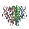 6c70MC M: atomic model generated by this map C: citing same article ( |
|---|---|
| Similar structure data |


-Links
| EMDB pages | EMDB (EBI/PDBe) / EMDataResource |
|---|
-Map
| File | Download / File: emd_7352.map.gz / Format: CCP4 / Size: 216 MB / Type: IMAGE STORED AS FLOATING POINT NUMBER (4 BYTES) | ||||||||||||||||||||||||||||||||||||||||||||||||||||||||||||
|---|---|---|---|---|---|---|---|---|---|---|---|---|---|---|---|---|---|---|---|---|---|---|---|---|---|---|---|---|---|---|---|---|---|---|---|---|---|---|---|---|---|---|---|---|---|---|---|---|---|---|---|---|---|---|---|---|---|---|---|---|---|
| Annotation | Odorant receptor | ||||||||||||||||||||||||||||||||||||||||||||||||||||||||||||
| Projections & slices | Image control
Images are generated by Spider. | ||||||||||||||||||||||||||||||||||||||||||||||||||||||||||||
| Voxel size | X=Y=Z: 1 Å | ||||||||||||||||||||||||||||||||||||||||||||||||||||||||||||
| Density |
| ||||||||||||||||||||||||||||||||||||||||||||||||||||||||||||
| Symmetry | Space group: 1 | ||||||||||||||||||||||||||||||||||||||||||||||||||||||||||||
| Details | EMDB XML:
CCP4 map header:
| ||||||||||||||||||||||||||||||||||||||||||||||||||||||||||||
 Z (Sec.)
 Y (Row.)
 X (Col.)


-Supplemental data
- Sample components
-Entire : Orco homotetramer bound to four Fab molecules in a detergent micelle
| Entire | Name: Orco homotetramer bound to four Fab molecules in a detergent micelle |
|---|---|
| Components |
|
-Supramolecule #1: Orco homotetramer bound to four Fab molecules in a detergent micelle
| Supramolecule | Name: Orco homotetramer bound to four Fab molecules in a detergent micelle type: complex / ID: 1 / Parent: 0 / Macromolecule list: all |
|---|---|
| Source (natural) | Organism: Apocrypta bakeri (insect) |
-Macromolecule #1: Odorant receptor
| Macromolecule | Name: Odorant receptor / type: protein_or_peptide / ID: 1 / Number of copies: 4 / Enantiomer: LEVO |
|---|---|
| Source (natural) | Organism: Apocrypta bakeri (insect) |
| Molecular weight | Theoretical: 53.513238 KDa |
| Recombinant expression | Organism:  Homo sapiens (human) Homo sapiens (human) |
| Sequence | String: GPGRAKFKHQ GLVADLLPNI RVMQGVGHFM FNYYSEGKKF PHRIYCIVTL LLLLLQYGMM AVNLMMESDD VDDLTANTIT MLFFLHPIV KMIYFPVRSK IFYKTLAIWN NPNSHPLFAE SNARFHALAI TKMRRLLFCV AGATIFSVIS WTGITFIEDS V KRITDPET ...String: GPGRAKFKHQ GLVADLLPNI RVMQGVGHFM FNYYSEGKKF PHRIYCIVTL LLLLLQYGMM AVNLMMESDD VDDLTANTIT MLFFLHPIV KMIYFPVRSK IFYKTLAIWN NPNSHPLFAE SNARFHALAI TKMRRLLFCV AGATIFSVIS WTGITFIEDS V KRITDPET NETTIIPIPR LMIRTFYPFN AMSGAGHVFA LIYQFYYLVI SMAVSNSLDV LFCSWLLFAC EQLQHLKAIM KP LMELSAT LDTVVPNSGE LFKAGSADHL RESQGVQPSG NGDNVLDVDL RGIYSNRQDF TATFRPTAGT TFNGGVGPNG LTK KQEMLV RSAIKYWVER HKHVVRLVTA VGDAYGVALL LHMLTTTITL TLLAYQATKV NGVNVYAATV IGYLLYTLGQ VFLF CIFGN RLIEESSSVM EAAYSCHWYD GSEEAKTFVQ IVCQQCQKAM SISGAKFFTV SLDLFASVLG AVVTYFMVLV QLK UniProtKB: Odorant receptor |
-Experimental details
-Structure determination
| Method | cryo EM |
|---|---|
 Processing Processing | single particle reconstruction |
| Aggregation state | particle |
-Sample preparation
| Buffer | pH: 7.5 |
|---|---|
| Vitrification | Cryogen name: ETHANE |
- Electron microscopy
| Microscope | FEI TITAN KRIOS |
|---|---|
| Image recording | Film or detector model: GATAN K2 SUMMIT (4k x 4k) / Detector mode: SUPER-RESOLUTION / Digitization - Frames/image: 1-50 / Number grids imaged: 1221 / Number real images: 53141 / Average exposure time: 0.2 sec. / Average electron dose: 80.0 e/Å2 |
| Electron beam | Acceleration voltage: 300 kV / Electron source:  FIELD EMISSION GUN FIELD EMISSION GUN |
| Electron optics | Illumination mode: FLOOD BEAM / Imaging mode: BRIGHT FIELD |
| Experimental equipment |  Model: Titan Krios / Image courtesy: FEI Company |
