 Movie
Movie Controller


+ Open data

- Basic information
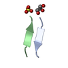
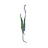
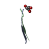
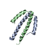
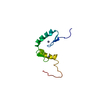
| Entry | Database: PDB / ID: 6c3f | ||||||
|---|---|---|---|---|---|---|---|
| Title | AMYLOID FORMING PEPTIDE IYKVEI FROM TRANSTHYRETIN | ||||||
 Components Components | ILE-TYR-LYS-VAL-GLU-ILE | ||||||
 Keywords Keywords | PROTEIN FIBRIL / amyloid / transthyretin / fibril | ||||||
| Function / homology | trifluoroacetic acid Function and homology information Function and homology information | ||||||
| Biological species |  Homo sapiens (human) Homo sapiens (human) | ||||||
| Method |  X-RAY DIFFRACTION / SYNCHROTRON / MOLECULAR REPLACEMENT / Resolution: 1.499 Å X-RAY DIFFRACTION / SYNCHROTRON / MOLECULAR REPLACEMENT / Resolution: 1.499 Å | ||||||
 Authors Authors | Saelices, L. / Sawaya, M.R. / Eisenberg, D.S. | ||||||
| Funding support |  United States, 1items United States, 1items
| ||||||
 Citation Citation | Journal: Protein Sci. / Year: 2018 Title: Crystal structures of amyloidogenic segments of human transthyretin. Authors: Saelices, L. / Sievers, S.A. / Sawaya, M.R. / Eisenberg, D.S. #1: Journal: J. Biol. Chem. / Year: 2015Title: Uncovering the Mechanism of Aggregation of Human Transthyretin. Authors: Saelices, L. / Johnson, L.M. / Liang, W.Y. / Sawaya, M.R. / Cascio, D. / Ruchala, P. / Whitelegge, J. / Jiang, L. / Riek, R. / Eisenberg, D.S. | ||||||
| History |
|
- Structure visualization
| Structure viewer | Molecule: MolmilJmol/JSmol |
|---|
- Downloads & links
-Download
| PDBx/mmCIF format | 6c3f.cif.gz | 12.4 KB | Display | PDBx/mmCIF format |
|---|---|---|---|---|
| PDB format | pdb6c3f.ent.gz | 6.2 KB | Display | PDB format |
| PDBx/mmJSON format | 6c3f.json.gz | Tree view | PDBx/mmJSON format | |
| Others |  Other downloads Other downloads |
-Validation report
| Arichive directory | https://data.pdbj.org/pub/pdb/validation_reports/c3/6c3fftp://data.pdbj.org/pub/pdb/validation_reports/c3/6c3f | HTTPS FTP |
|---|
-Related structure data


-Links
 PDBj
PDBj
- Assembly
| Deposited unit | 
| ||||||||
|---|---|---|---|---|---|---|---|---|---|
| 1 | x 10
| ||||||||
| Unit cell |
|
-Components
| #1: Protein/peptide | Mass: 764.929 Da / Num. of mol.: 2 / Source method: obtained synthetically / Source: (synth.) Homo sapiens (human)#2: Chemical | ChemComp-TFA / | 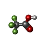  Mass: 114.023 Da / Num. of mol.: 1 / Source method: obtained synthetically / Formula: C2HF3O2 Mass: 114.023 Da / Num. of mol.: 1 / Source method: obtained synthetically / Formula: C2HF3O2#3: Water | ChemComp-HOH / |  Mass: 18.015 Da / Num. of mol.: 4 / Source method: isolated from a natural source / Formula: H2O Mass: 18.015 Da / Num. of mol.: 4 / Source method: isolated from a natural source / Formula: H2O |
|---|
-Experimental details
-Experiment
| Experiment | Method: X-RAY DIFFRACTION / Number of used crystals: 1 |
|---|
- Sample preparation
| Crystal | Density Matthews: 1.47 Å3/Da / Density % sol: 16.55 % |
|---|---|
| Crystal grow | Temperature: 298 K / Method: vapor diffusion, hanging drop / Details: 1.5 M NaCl, 10 % v/v ethanol, 25% Glycerol |
-Data collection
| Diffraction | Mean temperature: 100 K | ||||||||||||||||||||||||||||||||||||||||||||||||||||||||||||||||||||||||||||||||||||||||||||||||||||||||||||||||||||||||
|---|---|---|---|---|---|---|---|---|---|---|---|---|---|---|---|---|---|---|---|---|---|---|---|---|---|---|---|---|---|---|---|---|---|---|---|---|---|---|---|---|---|---|---|---|---|---|---|---|---|---|---|---|---|---|---|---|---|---|---|---|---|---|---|---|---|---|---|---|---|---|---|---|---|---|---|---|---|---|---|---|---|---|---|---|---|---|---|---|---|---|---|---|---|---|---|---|---|---|---|---|---|---|---|---|---|---|---|---|---|---|---|---|---|---|---|---|---|---|---|---|---|
| Diffraction source | Source: SYNCHROTRON / Site: APS / Beamline: 24-ID-E / Wavelength: 0.9792 Å | ||||||||||||||||||||||||||||||||||||||||||||||||||||||||||||||||||||||||||||||||||||||||||||||||||||||||||||||||||||||||
| Detector | Type: ADSC QUANTUM 315 / Detector: CCD / Date: Feb 28, 2014 | ||||||||||||||||||||||||||||||||||||||||||||||||||||||||||||||||||||||||||||||||||||||||||||||||||||||||||||||||||||||||
| Radiation | Protocol: SINGLE WAVELENGTH / Monochromatic (M) / Laue (L): M / Scattering type: x-ray | ||||||||||||||||||||||||||||||||||||||||||||||||||||||||||||||||||||||||||||||||||||||||||||||||||||||||||||||||||||||||
| Radiation wavelength | Wavelength: 0.9792 Å / Relative weight: 1 | ||||||||||||||||||||||||||||||||||||||||||||||||||||||||||||||||||||||||||||||||||||||||||||||||||||||||||||||||||||||||
| Reflection | Resolution: 1.499→15.952 Å / Num. obs: 1413 / % possible obs: 96.3 % / Redundancy: 2.735 % / Biso Wilson estimate: 5.02 Å2 / CC1/2: 0.989 / Rmerge(I) obs: 0.154 / Rrim(I) all: 0.188 / Χ2: 0.974 / Net I/σ(I): 5.06 | ||||||||||||||||||||||||||||||||||||||||||||||||||||||||||||||||||||||||||||||||||||||||||||||||||||||||||||||||||||||||
| Reflection shell | Diffraction-ID: 1
|
- Processing
| Software |
| ||||||||||||||||||||||||
|---|---|---|---|---|---|---|---|---|---|---|---|---|---|---|---|---|---|---|---|---|---|---|---|---|---|
| Refinement | Method to determine structure: MOLECULAR REPLACEMENT / Resolution: 1.499→15.952 Å / SU ML: 0.11 / Cross valid method: THROUGHOUT / σ(F): 1.39 / Phase error: 28.86
| ||||||||||||||||||||||||
| Solvent computation | Shrinkage radii: 0.9 Å / VDW probe radii: 1.11 Å | ||||||||||||||||||||||||
| Displacement parameters | Biso max: 17.89 Å2 / Biso mean: 8.12 Å2 / Biso min: 2.21 Å2 | ||||||||||||||||||||||||
| Refinement step | Cycle: final / Resolution: 1.499→15.952 Å
| ||||||||||||||||||||||||
| Refine LS restraints |
| ||||||||||||||||||||||||
| LS refinement shell | Resolution: 1.4991→1.553 Å / Rfactor Rfree error: 0 / Total num. of bins used: 1
|
