 Movie
Movie Controller

[English] 日本語
 Yorodumi
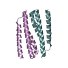
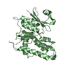
- PDB-4gqm: Crystal structure of a helix-turn-helix containing hypothetical p... -
+ Open data

- Basic information
| Entry | Database: PDB / ID: 4gqm | ||||||
|---|---|---|---|---|---|---|---|
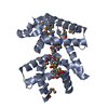
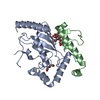
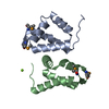
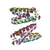
| Title | Crystal structure of a helix-turn-helix containing hypothetical protein (CT009) from Chlamydia trachomatis in a sub-domain swap conformation | ||||||
 Components Components | CT009 | ||||||
 Keywords Keywords | UNKNOWN FUNCTION / helix-turn-helix | ||||||
| Function / homology |  Function and homology information Function and homology information | ||||||
| Biological species |   Chlamydia trachomatis (bacteria) Chlamydia trachomatis (bacteria) | ||||||
| Method |  X-RAY DIFFRACTION / SYNCHROTRON / MOLECULAR REPLACEMENT / molecular replacement / Resolution: 1.25 Å X-RAY DIFFRACTION / SYNCHROTRON / MOLECULAR REPLACEMENT / molecular replacement / Resolution: 1.25 Å | ||||||
 Authors Authors | Kemege, K. / Hickey, J. / Lovell, S. / Battaile, K.P. / Hefty, P.S. | ||||||
 Citation Citation | Journal: To be Published Title: Crystal structure of a helix-turn-helix containing hypothetical protein (CT009) from Chlamydia trachomatis in a sub-domain swap conformation Authors: Kemege, K. / Hickey, J. / Lovell, S. / Battaile, K.P. / Hefty, P.S. | ||||||
| History |
|
- Structure visualization
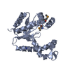
| Structure viewer | Molecule: MolmilJmol/JSmol |
|---|
- Downloads & links
-Download
| PDBx/mmCIF format | 4gqm.cif.gz | 65.3 KB | Display | PDBx/mmCIF format |
|---|---|---|---|---|
| PDB format | pdb4gqm.ent.gz | 49.4 KB | Display | PDB format |
| PDBx/mmJSON format | 4gqm.json.gz | Tree view | PDBx/mmJSON format | |
| Others |  Other downloads Other downloads |
-Validation report
| Arichive directory | https://data.pdbj.org/pub/pdb/validation_reports/gq/4gqmftp://data.pdbj.org/pub/pdb/validation_reports/gq/4gqm | HTTPS FTP |
|---|
-Related structure data
| Similar structure data |
|---|


-Links
 PDBj
PDBj


- Assembly
| Deposited unit | 
| ||||||||
|---|---|---|---|---|---|---|---|---|---|
| 1 | 
| ||||||||
| Unit cell |
|
-Components
| #1: Protein | Mass: 13391.338 Da / Num. of mol.: 1 / Fragment: UNP residues 1-116 Source method: isolated from a genetically manipulated source Source: (gene. exp.) Chlamydia trachomatis (bacteria) / Strain: L2/434/Bu / Gene: CTL0264 / Plasmid: pET21b / Production host: |
|---|---|
| #2: Water | ChemComp-HOH /  Mass: 18.015 Da / Num. of mol.: 111 / Source method: isolated from a natural source / Formula: H2O Mass: 18.015 Da / Num. of mol.: 111 / Source method: isolated from a natural source / Formula: H2O |
-Experimental details
-Experiment
| Experiment | Method: X-RAY DIFFRACTION / Number of used crystals: 1 |
|---|
- Sample preparation
| Crystal | Density Matthews: 2.5 Å3/Da / Density % sol: 50.87 % |
|---|---|
| Crystal grow | Temperature: 293 K / Method: vapor diffusion / pH: 8.5 Details: 30% w/v PEG4000, 0.1 M Tris, pH 8.5, 0.2 M magnesium chloride, VAPOR DIFFUSION, temperature 293K |
-Data collection
| Diffraction | Mean temperature: 100 K |
|---|---|
| Diffraction source | Source: SYNCHROTRON / Site: APS  / Beamline: 17-ID / Wavelength: 1 Å / Beamline: 17-ID / Wavelength: 1 Å |
| Detector | Type: DECTRIS PILATUS 6M / Detector: PIXEL / Date: Dec 3, 2011 |
| Radiation | Monochromator: Si(111) / Protocol: SINGLE WAVELENGTH / Monochromatic (M) / Laue (L): M / Scattering type: x-ray |
| Radiation wavelength | Wavelength: 1 Å / Relative weight: 1 |
| Reflection | Resolution: 1.25→46.762 Å / Num. obs: 38420 / % possible obs: 99.9 % / Observed criterion σ(F): 0 / Observed criterion σ(I): -3 / Redundancy: 8.3 % / Rmerge(I) obs: 0.036 / Net I/σ(I): 25.1 |
| Reflection shell | Resolution: 1.25→1.27 Å / Redundancy: 7.5 % / Rmerge(I) obs: 0.824 / Mean I/σ(I) obs: 2.5 / % possible all: 98.5 |
-Phasing
| Phasing | Method: molecular replacement |
|---|
- Processing
| Software |
| |||||||||||||||||||||||||||||||||||||||||||||||||||||||||||||||||||||||||||||||||||||||||||||||||||||||||
|---|---|---|---|---|---|---|---|---|---|---|---|---|---|---|---|---|---|---|---|---|---|---|---|---|---|---|---|---|---|---|---|---|---|---|---|---|---|---|---|---|---|---|---|---|---|---|---|---|---|---|---|---|---|---|---|---|---|---|---|---|---|---|---|---|---|---|---|---|---|---|---|---|---|---|---|---|---|---|---|---|---|---|---|---|---|---|---|---|---|---|---|---|---|---|---|---|---|---|---|---|---|---|---|---|---|---|
| Refinement | Method to determine structure: MOLECULAR REPLACEMENT Starting model: COMPUTATIONAL MODEL GENERATED FROM THE PROGRAM MODELLER Resolution: 1.25→29.208 Å / Occupancy max: 1 / Occupancy min: 0.16 / SU ML: 0.13 / Cross valid method: THROUGHOUT / σ(F): 1.91 / Phase error: 14.76 / Stereochemistry target values: ML
| |||||||||||||||||||||||||||||||||||||||||||||||||||||||||||||||||||||||||||||||||||||||||||||||||||||||||
| Solvent computation | Shrinkage radii: 1.2 Å / VDW probe radii: 1.3 Å / Solvent model: FLAT BULK SOLVENT MODEL | |||||||||||||||||||||||||||||||||||||||||||||||||||||||||||||||||||||||||||||||||||||||||||||||||||||||||
| Displacement parameters | Biso max: 49.78 Å2 / Biso mean: 20.238 Å2 / Biso min: 9.18 Å2 | |||||||||||||||||||||||||||||||||||||||||||||||||||||||||||||||||||||||||||||||||||||||||||||||||||||||||
| Refinement step | Cycle: LAST / Resolution: 1.25→29.208 Å
| |||||||||||||||||||||||||||||||||||||||||||||||||||||||||||||||||||||||||||||||||||||||||||||||||||||||||
| Refine LS restraints |
| |||||||||||||||||||||||||||||||||||||||||||||||||||||||||||||||||||||||||||||||||||||||||||||||||||||||||
| LS refinement shell | Refine-ID: X-RAY DIFFRACTION / Total num. of bins used: 14
|
