 Movie
Movie Controller

[English] 日本語
 Yorodumi
- PDB-4aeq: Crystal structure of the dimeric immunity protein Cmi solved by d... -
+ Open data

- Basic information
| Entry | Database: PDB / ID: 4aeq | ||||||
|---|---|---|---|---|---|---|---|
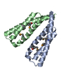
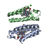
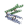
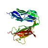
| Title | Crystal structure of the dimeric immunity protein Cmi solved by direct methods (Arcimboldo) | ||||||
 Components Components | COLICIN-M IMMUNITY PROTEIN | ||||||
 Keywords Keywords | IMMUNE SYSTEM / 3D DOMAIN SWAP | ||||||
| Function / homology | YebF/Colicin-M immunity protein / YebF/Cmi superfamily / YebF-like protein / YebF/Cmi domain profile. / bacteriocin immunity / PHOSPHATE ION / Colicin-M immunity protein Function and homology information Function and homology information | ||||||
| Biological species |  | ||||||
| Method |  X-RAY DIFFRACTION / SYNCHROTRON / DIRECT METHODS / Resolution: 1.892 Å X-RAY DIFFRACTION / SYNCHROTRON / DIRECT METHODS / Resolution: 1.892 Å | ||||||
 Authors Authors | Zeth, K. / Patzer, S.I. / Albrecht, R. / Uson, I. / Braun, V. | ||||||
 Citation Citation | Journal: J.Struct.Biol. / Year: 2012 Title: The Crystal Structure of the Dimeric Colicin M Immunity Authors: Uson, I. / Patzer, S.I. / Dayana Rodriguez, D.D. / Braun, V. / Zeth, K. | ||||||
| History |
|
- Structure visualization
| Structure viewer | Molecule: MolmilJmol/JSmol |
|---|
- Downloads & links
-Download
| PDBx/mmCIF format | 4aeq.cif.gz | 51.7 KB | Display | PDBx/mmCIF format |
|---|---|---|---|---|
| PDB format | pdb4aeq.ent.gz | 37.1 KB | Display | PDB format |
| PDBx/mmJSON format | 4aeq.json.gz | Tree view | PDBx/mmJSON format | |
| Others |  Other downloads Other downloads |
-Validation report
| Arichive directory | https://data.pdbj.org/pub/pdb/validation_reports/ae/4aeqftp://data.pdbj.org/pub/pdb/validation_reports/ae/4aeq | HTTPS FTP |
|---|
-Related structure data
| Related structure data | |
|---|---|
| Similar structure data |


-Links
 PDBj
PDBj- Assembly
| Deposited unit | 
| ||||||||
|---|---|---|---|---|---|---|---|---|---|
| 1 | 
| ||||||||
| Unit cell |
| ||||||||
| Components on special symmetry positions |
|
-Components
| #1: Protein | Mass: 11670.021 Da / Num. of mol.: 1 / Fragment: RESIDUES 44-141 Source method: isolated from a genetically manipulated source Details: CMI CLONED WITHOUT THE N-TERMINAL HYDROPHOBIC HELIX FOR EXPRESSION OF THE PROTEIN IN THE CYTOPLASM Source: (gene. exp.) |
|---|---|
| #2: Chemical | ChemComp-PO4 /   Mass: 94.971 Da / Num. of mol.: 1 / Source method: obtained synthetically / Formula: PO4 Mass: 94.971 Da / Num. of mol.: 1 / Source method: obtained synthetically / Formula: PO4 |
| #3: Water | ChemComp-HOH /  Mass: 18.015 Da / Num. of mol.: 54 / Source method: isolated from a natural source / Formula: H2O Mass: 18.015 Da / Num. of mol.: 54 / Source method: isolated from a natural source / Formula: H2O |
| Has protein modification | Y |
-Experimental details
-Experiment
| Experiment | Method: X-RAY DIFFRACTION / Number of used crystals: 1 |
|---|
- Sample preparation
| Crystal | Density Matthews: 2.26 Å3/Da / Density % sol: 45.65 % / Description: NONE |
|---|---|
| Crystal grow | Details: 0.2 M AMMONIUM SULFATE, 30% PEG 4000 |
-Data collection
| Diffraction | Mean temperature: 100 K |
|---|---|
| Diffraction source | Source: SYNCHROTRON / Site: SLS  / Beamline: X10SA / Wavelength: 1 / Beamline: X10SA / Wavelength: 1 |
| Detector | Type: DECTRIS PILATUS 6M / Detector: PIXEL |
| Radiation | Protocol: SINGLE WAVELENGTH / Monochromatic (M) / Laue (L): M / Scattering type: x-ray |
| Radiation wavelength | Wavelength: 1 Å / Relative weight: 1 |
| Reflection | Resolution: 1.89→50 Å / Num. obs: 15884 / % possible obs: 96.6 % / Observed criterion σ(I): 2.5 / Redundancy: 2.6 % / Biso Wilson estimate: 23.36 Å2 / Rmerge(I) obs: 0.08 / Net I/σ(I): 11 |
| Reflection shell | Resolution: 1.89→2 Å / Redundancy: 2.5 % / Rmerge(I) obs: 0.51 / Mean I/σ(I) obs: 2.5 / % possible all: 90 |
- Processing
| Software |
| |||||||||||||||||||||||||||||||||||||||||||||||||||||||||||||||||||||||||||
|---|---|---|---|---|---|---|---|---|---|---|---|---|---|---|---|---|---|---|---|---|---|---|---|---|---|---|---|---|---|---|---|---|---|---|---|---|---|---|---|---|---|---|---|---|---|---|---|---|---|---|---|---|---|---|---|---|---|---|---|---|---|---|---|---|---|---|---|---|---|---|---|---|---|---|---|---|
| Refinement | Method to determine structure: DIRECT METHODS Starting model: ALPHA HELIX Resolution: 1.892→28.224 Å / SU ML: 0.22 / σ(F): 2.03 / Phase error: 23.05 / Stereochemistry target values: ML
| |||||||||||||||||||||||||||||||||||||||||||||||||||||||||||||||||||||||||||
| Solvent computation | Shrinkage radii: 0.49 Å / VDW probe radii: 0.8 Å / Solvent model: FLAT BULK SOLVENT MODEL / Bsol: 63.597 Å2 / ksol: 0.455 e/Å3 | |||||||||||||||||||||||||||||||||||||||||||||||||||||||||||||||||||||||||||
| Displacement parameters |
| |||||||||||||||||||||||||||||||||||||||||||||||||||||||||||||||||||||||||||
| Refinement step | Cycle: LAST / Resolution: 1.892→28.224 Å
| |||||||||||||||||||||||||||||||||||||||||||||||||||||||||||||||||||||||||||
| Refine LS restraints |
| |||||||||||||||||||||||||||||||||||||||||||||||||||||||||||||||||||||||||||
| LS refinement shell |
| |||||||||||||||||||||||||||||||||||||||||||||||||||||||||||||||||||||||||||
| Refinement TLS params. | Method: refined / Refine-ID: X-RAY DIFFRACTION
| |||||||||||||||||||||||||||||||||||||||||||||||||||||||||||||||||||||||||||
| Refinement TLS group |
|
