 ムービー
ムービー コントローラー


+ データを開く

- 基本情報
基本情報
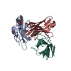
| 登録情報 | データベース: PDB / ID: 6c10 | ||||||
|---|---|---|---|---|---|---|---|
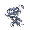
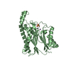
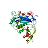
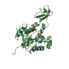
| タイトル | Crystal structure of mouse PCDH15 EC11-EL | ||||||
 要素 要素 | Protocadherin-15 | ||||||
 キーワード キーワード | membrane protein / metal transport / PCDH15 / LHFPL5 / protocadherin / tip link / hair cell / TMHS / hearing | ||||||
| 機能・相同性 |  機能・相同性情報 機能・相同性情報detection of mechanical stimulus involved in equilibrioception / inner ear receptor cell stereocilium organization / inner ear auditory receptor cell differentiation / righting reflex / stereocilium bundle / detection of mechanical stimulus involved in sensory perception of sound / stereocilium / non-motile cilium assembly / auditory receptor cell stereocilium organization / adult walking behavior ...detection of mechanical stimulus involved in equilibrioception / inner ear receptor cell stereocilium organization / inner ear auditory receptor cell differentiation / righting reflex / stereocilium bundle / detection of mechanical stimulus involved in sensory perception of sound / stereocilium / non-motile cilium assembly / auditory receptor cell stereocilium organization / adult walking behavior / startle response / homophilic cell adhesion via plasma membrane adhesion molecules / inner ear development / actin filament bundle assembly / photoreceptor outer segment / visual perception / actin filament organization / morphogenesis of an epithelium / locomotory behavior / sensory perception of sound / response to calcium ion / multicellular organism growth / calcium ion binding / extracellular region / membrane / plasma membrane / cytoplasm 類似検索 - 分子機能 | ||||||
| 生物種 |  | ||||||
| 手法 |  X線回折 / シンクロトロン / 分子置換 / 解像度: 1.399 Å X線回折 / シンクロトロン / 分子置換 / 解像度: 1.399 Å | ||||||
 データ登録者 データ登録者 | Gouaux, E. / Elferich, J. / Ge, J. | ||||||
 引用 引用 | ジャーナル: Elife / 年: 2018 タイトル: Structure of mouse protocadherin 15 of the stereocilia tip link in complex with LHFPL5. 著者: Jingpeng Ge / Johannes Elferich / April Goehring / Huaying Zhao / Peter Schuck / Eric Gouaux /  要旨: Hearing and balance involve the transduction of mechanical stimuli into electrical signals by deflection of bundles of stereocilia linked together by protocadherin 15 (PCDH15) and cadherin 23 'tip ...Hearing and balance involve the transduction of mechanical stimuli into electrical signals by deflection of bundles of stereocilia linked together by protocadherin 15 (PCDH15) and cadherin 23 'tip links'. PCDH15 transduces tip link tension into opening of a mechano-electrical transduction (MET) ion channel. PCDH15 also interacts with LHFPL5, a candidate subunit of the MET channel. Here we illuminate the PCDH15-LHFPL5 structure, showing how the complex is composed of PCDH15 and LHFPL5 subunit pairs related by a 2-fold axis. The extracellular cadherin domains define a mobile tether coupled to a rigid, 2-fold symmetric 'collar' proximal to the membrane bilayer. LHFPL5 forms extensive interactions with the PCDH15 transmembrane helices and stabilizes the overall PCDH15-LHFPL5 assembly. Our studies illuminate the architecture of the PCDH15-LHFPL5 complex, localize mutations associated with deafness, and shed new light on how forces in the PCDH15 tether may be transduced into the stereocilia membrane. | ||||||
| 履歴 |
|
- 構造の表示
構造の表示
| 構造ビューア | 分子: MolmilJmol/JSmol |
|---|
- ダウンロードとリンク
-ダウンロード
| PDBx/mmCIF形式 | 6c10.cif.gz | 116 KB | 表示 | PDBx/mmCIF形式 |
|---|---|---|---|---|
| PDB形式 | pdb6c10.ent.gz | 88.1 KB | 表示 | PDB形式 |
| PDBx/mmJSON形式 | 6c10.json.gz | ツリー表示 | PDBx/mmJSON形式 | |
| その他 |  その他のダウンロード その他のダウンロード |
-検証レポート
| 文書・要旨 | 6c10_validation.pdf.gz | 441.1 KB | 表示 | wwPDB検証レポート |
|---|---|---|---|---|
| 文書・詳細版 | 6c10_full_validation.pdf.gz | 443.3 KB | 表示 | |
| XML形式データ | 6c10_validation.xml.gz | 14.4 KB | 表示 | |
| CIF形式データ | 6c10_validation.cif.gz | 19.6 KB | 表示 | |
| アーカイブディレクトリ | https://data.pdbj.org/pub/pdb/validation_reports/c1/6c10ftp://data.pdbj.org/pub/pdb/validation_reports/c1/6c10 | HTTPS FTP |
-関連構造データ


-リンク
 PDBj
PDBj


- 集合体
集合体
| 登録構造単位 | 
| ||||||||
|---|---|---|---|---|---|---|---|---|---|
| 1 |
| ||||||||
| 単位格子 |
|
-要素
| #1: タンパク質 | 分子量: 27911.832 Da / 分子数: 1 / 由来タイプ: 組換発現 / 由来: (組換発現)  Homo sapiens (ヒト) / 参照: UniProt: Q99PJ1 Homo sapiens (ヒト) / 参照: UniProt: Q99PJ1 |
|---|---|
| #2: 糖 | ChemComp-MAN /   タイプ: D-saccharide, alpha linking / 分子量: 180.156 Da / 分子数: 1 / 由来タイプ: 組換発現 / 式: C6H12O6 タイプ: D-saccharide, alpha linking / 分子量: 180.156 Da / 分子数: 1 / 由来タイプ: 組換発現 / 式: C6H12O6 |
| #3: 水 | ChemComp-HOH /  分子量: 18.015 Da / 分子数: 187 / 由来タイプ: 天然 / 式: H2O 分子量: 18.015 Da / 分子数: 187 / 由来タイプ: 天然 / 式: H2O |
| Has protein modification | Y |
-実験情報
-実験
| 実験 | 手法: X線回折 / 使用した結晶の数: 1 |
|---|
- 試料調製
試料調製
| 結晶 | マシュー密度: 2.37 Å3/Da / 溶媒含有率: 48.14 % |
|---|---|
| 結晶化 | 温度: 293 K / 手法: 蒸気拡散法, ハンギングドロップ法 / pH: 8.5 詳細: 0.2 M Lithium sulfate, 30% (w/v) PEG4000, 0.1 M Tris pH 8.5 |
-データ収集
| 回折 | 平均測定温度: 100 K |
|---|---|
| 放射光源 | 由来: シンクロトロン / サイト: APS / ビームライン: 24-ID-C / 波長: 0.98 Å |
| 検出器 | タイプ: DECTRIS PILATUS 6M-F / 検出器: PIXEL / 日付: 2017年6月18日 |
| 放射 | プロトコル: SINGLE WAVELENGTH / 単色(M)・ラウエ(L): M / 散乱光タイプ: x-ray |
| 放射波長 | 波長: 0.98 Å / 相対比: 1 |
| 反射 | 解像度: 1.4→54.23 Å / Num. obs: 53914 / % possible obs: 99.73 % / 冗長度: 9.7 % / Net I/σ(I): 25.05 |
| 反射 シェル | 解像度: 1.4→1.45 Å |
- 解析
解析
| ソフトウェア |
| |||||||||||||||||||||||||||||||||||||||||||||||||||||||||||||||||||||||||||||||||||||||||||||||||||||||||||||||||||||||||||||||||||||||||||||||||||||||||||||||||||||||||||||||||||||||||||||||||||||||||||||||||||||||||
|---|---|---|---|---|---|---|---|---|---|---|---|---|---|---|---|---|---|---|---|---|---|---|---|---|---|---|---|---|---|---|---|---|---|---|---|---|---|---|---|---|---|---|---|---|---|---|---|---|---|---|---|---|---|---|---|---|---|---|---|---|---|---|---|---|---|---|---|---|---|---|---|---|---|---|---|---|---|---|---|---|---|---|---|---|---|---|---|---|---|---|---|---|---|---|---|---|---|---|---|---|---|---|---|---|---|---|---|---|---|---|---|---|---|---|---|---|---|---|---|---|---|---|---|---|---|---|---|---|---|---|---|---|---|---|---|---|---|---|---|---|---|---|---|---|---|---|---|---|---|---|---|---|---|---|---|---|---|---|---|---|---|---|---|---|---|---|---|---|---|---|---|---|---|---|---|---|---|---|---|---|---|---|---|---|---|---|---|---|---|---|---|---|---|---|---|---|---|---|---|---|---|---|---|---|---|---|---|---|---|---|---|---|---|---|---|---|---|---|
| 精密化 | 構造決定の手法: 分子置換 / 解像度: 1.399→54.23 Å / SU ML: 0.17 / 交差検証法: FREE R-VALUE / σ(F): 1.32 / 位相誤差: 21.02 / 立体化学のターゲット値: ML
| |||||||||||||||||||||||||||||||||||||||||||||||||||||||||||||||||||||||||||||||||||||||||||||||||||||||||||||||||||||||||||||||||||||||||||||||||||||||||||||||||||||||||||||||||||||||||||||||||||||||||||||||||||||||||
| 溶媒の処理 | 減衰半径: 0.9 Å / VDWプローブ半径: 1.11 Å / 溶媒モデル: FLAT BULK SOLVENT MODEL | |||||||||||||||||||||||||||||||||||||||||||||||||||||||||||||||||||||||||||||||||||||||||||||||||||||||||||||||||||||||||||||||||||||||||||||||||||||||||||||||||||||||||||||||||||||||||||||||||||||||||||||||||||||||||
| 精密化ステップ | サイクル: LAST / 解像度: 1.399→54.23 Å
| |||||||||||||||||||||||||||||||||||||||||||||||||||||||||||||||||||||||||||||||||||||||||||||||||||||||||||||||||||||||||||||||||||||||||||||||||||||||||||||||||||||||||||||||||||||||||||||||||||||||||||||||||||||||||
| 拘束条件 |
| |||||||||||||||||||||||||||||||||||||||||||||||||||||||||||||||||||||||||||||||||||||||||||||||||||||||||||||||||||||||||||||||||||||||||||||||||||||||||||||||||||||||||||||||||||||||||||||||||||||||||||||||||||||||||
| LS精密化 シェル |
|
