National Institutes of Health/Office of the Director
AG-04182
米国
National Science Foundation (NSF, United States)
MCB-0958111
米国
引用
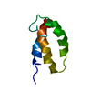
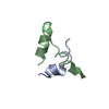
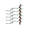
ジャーナル: Science / 年: 2018 タイトル: Atomic structures of low-complexity protein segments reveal kinked β sheets that assemble networks. 著者: Michael P Hughes / Michael R Sawaya / David R Boyer / Lukasz Goldschmidt / Jose A Rodriguez / Duilio Cascio / Lisa Chong / Tamir Gonen / David S Eisenberg / 要旨: Subcellular membraneless assemblies are a reinvigorated area of study in biology, with spirited scientific discussions on the forces between the low-complexity protein domains within these assemblies. ...Subcellular membraneless assemblies are a reinvigorated area of study in biology, with spirited scientific discussions on the forces between the low-complexity protein domains within these assemblies. To illuminate these forces, we determined the atomic structures of five segments from protein low-complexity domains associated with membraneless assemblies. Their common structural feature is the stacking of segments into kinked β sheets that pair into protofilaments. Unlike steric zippers of amyloid fibrils, the kinked sheets interact weakly through polar atoms and aromatic side chains. By computationally threading the human proteome on our kinked structures, we identified hundreds of low-complexity segments potentially capable of forming such interactions. These segments are found in proteins as diverse as RNA binders, nuclear pore proteins, and keratins, which are known to form networks and localize to membraneless assemblies.
履歴
登録
2017年12月25日
登録サイト: RCSB / 処理サイト: RCSB
改定 1.0
2018年4月4日
Provider: repository / タイプ: Initial release
改定 1.1
2018年4月25日
Group: Data collection / カテゴリ: diffrn_source / Item: _diffrn_source.source
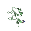
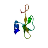
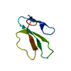
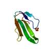
名称: A fibril composed of a 6-residue segment of FUS / タイプ: COMPLEX / Entity ID: #1 / 由来: NATURAL
分子量
実験値: NO
由来(天然)
生物種: Homo sapiens (ヒト)
EM crystal formation
装置: 24-well plate Atmosphere: air, sealed chaomder, in equilibrium with reservoir solutionq 詳細: 1 microliter of a 150 mg/mL peptide solution of STGGYG in water was mixed with 1 microliter of reservoir solution. The tray was incubated at room temperature and crystals grew within a week. Lipid mixture: none / 温度: 298 K / Time: 1 DAY
緩衝液
pH: 4.6
緩衝液成分
ID
濃度
名称
式
Buffer-ID
1
0.1M
sodiumacetate
C2H3NaO2
1
2
0.15M
ammoniumsulfate
(NH4)2SO4
1
3
25 (w/v)
PEG2000MME
1
試料
濃度: 150 mg/ml / 包埋: NO / シャドウイング: NO / 染色: NO / 凍結: YES / 詳細: crystal
凍結剤: NITROGEN 試料ホルダーモデル: GATAN 626 SINGLE TILT LIQUID NITROGEN CRYO TRANSFER HOLDER 最高温度: 100 K / 最低温度: 100 K
撮影
平均露光時間: 2 sec. / 電子線照射量: 0.01 e/Å2 フィルム・検出器のモデル: TVIPS TEMCAM-F416 (4k x 4k) 撮影したグリッド数: 2 詳細: The detector was operated in rolling shutter with 2x2 pixel binning.
画像スキャン
サンプリングサイズ: 15.6 µm / 横: 4096 / 縦: 4096
EM回折
カメラ長: 1350 mm
EM回折 シェル
解像度 (Å)
ID
EM diffraction stats-ID
フーリエ空間範囲 (%)
多重度
構造因子数
位相残差 (°)
1.3861-12.778
1
1
99.2
8.3
1742
37.98
1.1004-1.3861
2
1
91.6
5.9
1476
32.15
EM回折 統計
詳細: Phase statistics are not applicable. No imaging was used. THe phases were obtained by a crystalloghraphic direct methods program, SHELXD. フーリエ空間範囲: 0.955 % / 再高解像度: 1.1 Å / 測定した強度の数: 23271 / 構造因子数: 3220 / 位相誤差: 35.3 ° / 位相残差: 0.1 ° / 位相誤差の除外基準: 0 / Rmerge: 0.266 / Rsym: 0.25
回折
平均測定温度: 100 K
放射光源
由来: ELECTRON MICROSCOPE / タイプ: TECNAI F20 TEM / 波長: 0.0251 Å
∠α: 90 ° / ∠β: 90 ° / ∠γ: 90 ° / A: 13.79 Å / B: 4.93 Å / C: 101.9 Å / 空間群名: P212121 / 空間群番号: 19
CTF補正
タイプ: NONE
3次元再構成
解像度: 1.1 Å / 解像度の算出法: DIFFRACTION PATTERN/LAYERLINES 詳細: Density map was obtained using measured diffration intensities and phases acquired from a crystallographic direct methods program, SHELXD. 対称性のタイプ: 3D CRYSTAL
原子モデル構築
B value: 8.09 / プロトコル: OTHER / 空間: RECIPROCAL / Target criteria: maximum liklihood
精密化
構造決定の手法: AB INITIO PHASING / 解像度: 1.1→13.31 Å / Cor.coef. Fo:Fc: 0.916 / Cor.coef. Fo:Fc free: 0.92 / SU R Cruickshank DPI: 0.045 / 交差検証法: THROUGHOUT / σ(F): 0 / SU R Blow DPI: 0.048 / SU Rfree Blow DPI: 0.051 / SU Rfree Cruickshank DPI: 0.048
 ムービー
ムービー コントローラー


 データを開く
 基本情報
基本情報 要素
要素 キーワード
キーワード 機能・相同性情報
 Homo sapiens (ヒト)
 データ登録者
 米国, 3件
  引用
引用 構造の表示
構造の表示 ダウンロードとリンク
 その他のダウンロード
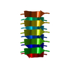


 PDBj
PDBj


 集合体
集合体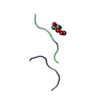

 分子量: 164.200 Da / 分子数: 1 / 断片: residues 77-82 / 由来タイプ: 合成 / 式: C7H16O4
 分子量: 18.015 Da / 分子数: 3 / 由来タイプ: 天然 / 式: H2O
 試料調製
試料調製
 FIELD EMISSION GUN / 加速電圧: 200 kV / 照射モード: FLOOD BEAM
 解析
解析