Entry Database : PDB / ID : 6bxzTitle Crystal Structure of Pig Protocadherin-15 EC10-MAD12 Protocadherin related 15 Keywords / / / / Function / homology Function Domain/homology Component
/ / / / / / / / / / / / / / / / / / / / / Biological species Sus scrofa (pig)Method / / / Resolution : 2.09 Å Authors De-la-Torre, P. / Araya-Secchi, R. / Choudhary, D. / Sotomayor, M. Funding support Organization Grant number Country National Institutes of Health/National Institute on Deafness and Other Communication Disorders (NIH/NIDCD) R01 DC015271 National Institutes of Health/National Institute on Deafness and Other Communication Disorders (NIH/NIDCD) R00 DC012534
Journal : Biophys. J. / Year : 2018Title : A Mechanically Weak Extracellular Membrane-Adjacent Domain Induces Dimerization of Protocadherin-15.Authors : De-la-Torre, P. / Choudhary, D. / Araya-Secchi, R. / Narui, Y. / Sotomayor, M. History Deposition Dec 19, 2017 Deposition site / Processing site Revision 1.0 Nov 28, 2018 Provider / Type Revision 1.1 Dec 26, 2018 Group / Database references / Category Item / _citation.pdbx_database_id_PubMed / _citation.titleRevision 1.2 Jan 2, 2019 Group / Database references / Category Item / _citation.page_first / _citation.page_lastRevision 1.3 Dec 11, 2019 Group / Category / Item Revision 1.4 Oct 4, 2023 Group Data collection / Database references ... Data collection / Database references / Derived calculations / Refinement description Category chem_comp_atom / chem_comp_bond ... chem_comp_atom / chem_comp_bond / database_2 / pdbx_initial_refinement_model / struct_conn / struct_ncs_dom_lim Item _database_2.pdbx_DOI / _database_2.pdbx_database_accession ... _database_2.pdbx_DOI / _database_2.pdbx_database_accession / _struct_conn.pdbx_dist_value / _struct_conn.ptnr1_auth_asym_id / _struct_conn.ptnr1_auth_comp_id / _struct_conn.ptnr1_auth_seq_id / _struct_conn.ptnr1_label_asym_id / _struct_conn.ptnr1_label_atom_id / _struct_conn.ptnr1_label_comp_id / _struct_conn.ptnr1_label_seq_id / _struct_conn.ptnr2_auth_asym_id / _struct_conn.ptnr2_auth_comp_id / _struct_conn.ptnr2_auth_seq_id / _struct_conn.ptnr2_label_asym_id / _struct_conn.ptnr2_label_atom_id / _struct_conn.ptnr2_label_comp_id / _struct_ncs_dom_lim.beg_auth_comp_id / _struct_ncs_dom_lim.beg_label_asym_id / _struct_ncs_dom_lim.beg_label_comp_id / _struct_ncs_dom_lim.beg_label_seq_id / _struct_ncs_dom_lim.end_auth_comp_id / _struct_ncs_dom_lim.end_label_asym_id / _struct_ncs_dom_lim.end_label_comp_id / _struct_ncs_dom_lim.end_label_seq_id
Show all Show less
 Movie
Movie Controller


 Open data
 Basic information
 Components
 Keywords
 Function and homology information
 X-RAY DIFFRACTION /
  Authors
 United States, 2items
  Citation
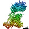
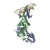
 Structure visualization
 Downloads & links
 Other downloads
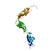

 PDBj
PDBj
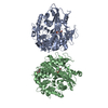
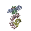
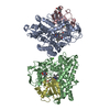
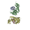
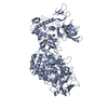
 Assembly


 Mass: 40.078 Da / Num. of mol.: 6 / Source method: obtained synthetically / Formula: Ca
 Mass: 18.015 Da / Num. of mol.: 260 / Source method: isolated from a natural source / Formula: H2O
 Sample preparation
 Processing