 Movie
Movie Controller


+ Open data

- Basic information
| Entry | Database: PDB / ID: 6bvb | ||||||||||||
|---|---|---|---|---|---|---|---|---|---|---|---|---|---|
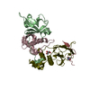
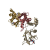
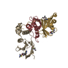
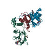
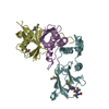
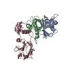
| Title | Crystal structure of HIF-2alpha-pVHL-elongin B-elongin C | ||||||||||||
 Components Components |
| ||||||||||||
 Keywords Keywords | GENE REGULATION / E3 Ubiquitin Ligase | ||||||||||||
| Function / homology |  Function and homology information Function and homology informationmyoblast fate commitment / Cellular response to hypoxia / PTK6 Expression / Transcriptional regulation of pluripotent stem cells / regulation of protein neddylation / regulation of cellular response to hypoxia / intracellular oxygen homeostasis / norepinephrine metabolic process / negative regulation of receptor signaling pathway via JAK-STAT / RHOBTB3 ATPase cycle ...myoblast fate commitment / Cellular response to hypoxia / PTK6 Expression / Transcriptional regulation of pluripotent stem cells / regulation of protein neddylation / regulation of cellular response to hypoxia / intracellular oxygen homeostasis / norepinephrine metabolic process / negative regulation of receptor signaling pathway via JAK-STAT / RHOBTB3 ATPase cycle / surfactant homeostasis / target-directed miRNA degradation / elongin complex / epithelial cell maturation / Replication of the SARS-CoV-1 genome / transcription elongation factor activity / Regulation of gene expression by Hypoxia-inducible Factor / VCB complex / Cul5-RING ubiquitin ligase complex / ubiquitin-dependent protein catabolic process via the C-end degron rule pathway / Cul2-RING ubiquitin ligase complex / SUMOylation of ubiquitinylation proteins / negative regulation of transcription elongation by RNA polymerase II / Pausing and recovery of Tat-mediated HIV elongation / Tat-mediated HIV elongation arrest and recovery / HIV elongation arrest and recovery / Pausing and recovery of HIV elongation / blood vessel remodeling / embryonic placenta development / negative regulation of signal transduction / Tat-mediated elongation of the HIV-1 transcript / Formation of HIV-1 elongation complex containing HIV-1 Tat / Formation of HIV elongation complex in the absence of HIV Tat / ubiquitin-like ligase-substrate adaptor activity / RNA Polymerase II Transcription Elongation / Formation of RNA Pol II elongation complex / negative regulation of TORC1 signaling / RNA Polymerase II Pre-transcription Events / visual perception / lung development / regulation of heart rate / ciliary tip / Pexophagy / transcription corepressor binding / negative regulation of autophagy / protein serine/threonine kinase binding / erythrocyte differentiation / RNA polymerase II transcription regulatory region sequence-specific DNA binding / TP53 Regulates Transcription of DNA Repair Genes / positive regulation of cell differentiation / transcription initiation at RNA polymerase II promoter / mitochondrion organization / transcription elongation by RNA polymerase II / Vif-mediated degradation of APOBEC3G / Inactivation of CSF3 (G-CSF) signaling / multicellular organismal-level iron ion homeostasis / mRNA transcription by RNA polymerase II / Evasion by RSV of host interferon responses / Oxygen-dependent proline hydroxylation of Hypoxia-inducible Factor Alpha / transcription coactivator binding / cell morphogenesis / Regulation of expression of SLITs and ROBOs / ubiquitin-protein transferase activity / transcription corepressor activity / Antigen processing: Ubiquitination & Proteasome degradation / positive regulation of proteasomal ubiquitin-dependent protein catabolic process / positive regulation of cold-induced thermogenesis / microtubule cytoskeleton / Neddylation / regulation of gene expression / protein-containing complex assembly / response to oxidative stress / angiogenesis / DNA-binding transcription activator activity, RNA polymerase II-specific / Replication of the SARS-CoV-2 genome / transcription regulator complex / cellular response to hypoxia / DNA-binding transcription factor binding / amyloid fibril formation / molecular adaptor activity / ubiquitin-dependent protein catabolic process / RNA polymerase II-specific DNA-binding transcription factor binding / proteasome-mediated ubiquitin-dependent protein catabolic process / protein-macromolecule adaptor activity / DNA-binding transcription factor activity, RNA polymerase II-specific / response to hypoxia / nuclear speck / protein stabilization / cilium / RNA polymerase II cis-regulatory region sequence-specific DNA binding / protein ubiquitination / protein heterodimerization activity / negative regulation of cell population proliferation / negative regulation of gene expression / ubiquitin protein ligase binding / regulation of transcription by RNA polymerase II / regulation of DNA-templated transcription / negative regulation of apoptotic process / positive regulation of DNA-templated transcription / chromatin Similarity search - Function | ||||||||||||
| Biological species |  Homo sapiens (human) Homo sapiens (human) | ||||||||||||
| Method |  X-RAY DIFFRACTION / SYNCHROTRON / MOLECULAR REPLACEMENT / Resolution: 2.002 Å X-RAY DIFFRACTION / SYNCHROTRON / MOLECULAR REPLACEMENT / Resolution: 2.002 Å | ||||||||||||
 Authors Authors | Tarade, D. / Ohh, M. / Lee, J.E. | ||||||||||||
| Funding support |  Canada, 3items Canada, 3items
| ||||||||||||
 Citation Citation | Journal: Nat Commun / Year: 2018 Title: HIF-2 alpha-pVHL complex reveals broad genotype-phenotype correlations in HIF-2 alpha-driven disease. Authors: Tarade, D. / Robinson, C.M. / Lee, J.E. / Ohh, M. | ||||||||||||
| History |
|
- Structure visualization
| Structure viewer | Molecule: MolmilJmol/JSmol |
|---|
- Downloads & links
-Download
| PDBx/mmCIF format | 6bvb.cif.gz | 217.7 KB | Display | PDBx/mmCIF format |
|---|---|---|---|---|
| PDB format | pdb6bvb.ent.gz | 177.8 KB | Display | PDB format |
| PDBx/mmJSON format | 6bvb.json.gz | Tree view | PDBx/mmJSON format | |
| Others |  Other downloads Other downloads |
-Validation report
| Arichive directory | https://data.pdbj.org/pub/pdb/validation_reports/bv/6bvbftp://data.pdbj.org/pub/pdb/validation_reports/bv/6bvb | HTTPS FTP |
|---|
-Related structure data
| Related structure data |  1lm8S S: Starting model for refinement |
|---|---|
| Similar structure data |


-Links
 PDBj
PDBj


- Assembly
| Deposited unit | 
| ||||||||
|---|---|---|---|---|---|---|---|---|---|
| 1 |
| ||||||||
| Unit cell |
|
-Components
| #1: Protein | Mass: 18702.291 Da / Num. of mol.: 1 Source method: isolated from a genetically manipulated source Source: (gene. exp.) Homo sapiens (human) / Gene: VHL / Production host:  |
|---|---|
| #2: Protein | Mass: 13147.781 Da / Num. of mol.: 1 Source method: isolated from a genetically manipulated source Source: (gene. exp.) Homo sapiens (human) / Gene: ELOB, TCEB2 / Production host: |
| #3: Protein | Mass: 10843.420 Da / Num. of mol.: 1 Source method: isolated from a genetically manipulated source Source: (gene. exp.) Homo sapiens (human) / Gene: ELOC, TCEB1 / Production host: |
| #4: Protein/peptide | Mass: 2197.416 Da / Num. of mol.: 1 / Source method: obtained synthetically / Source: (synth.) Homo sapiens (human) / References: UniProt: Q99814*PLUS |
| #5: Water | ChemComp-HOH /  Mass: 18.015 Da / Num. of mol.: 102 / Source method: isolated from a natural source / Formula: H2O Mass: 18.015 Da / Num. of mol.: 102 / Source method: isolated from a natural source / Formula: H2O |
-Experimental details
-Experiment
| Experiment | Method: X-RAY DIFFRACTION / Number of used crystals: 1 |
|---|
- Sample preparation
| Crystal | Density Matthews: 2.45 Å3/Da / Density % sol: 49.76 % / Description: Trapezoidal plates. |
|---|---|
| Crystal grow | Temperature: 293 K / Method: vapor diffusion, sitting drop / pH: 6.5 Details: 26% (v/v) PEG-3350, 0.2 M Li2SO4, 0.1 M Bis-Tris. 7 mg/mL protein complex. Temp details: Drops were set at 277 K. Incubated at 293 K. |
-Data collection
| Diffraction | Mean temperature: 100 K |
|---|---|
| Diffraction source | Source: SYNCHROTRON / Site: CLSI / Beamline: 08ID-1 / Wavelength: 1.28339 Å |
| Detector | Type: DECTRIS PILATUS 6M / Detector: PIXEL / Date: Sep 10, 2017 |
| Radiation | Protocol: SINGLE WAVELENGTH / Monochromatic (M) / Laue (L): M / Scattering type: x-ray |
| Radiation wavelength | Wavelength: 1.28339 Å / Relative weight: 1 |
| Reflection | Resolution: 2.002→48.307 Å / Num. obs: 31053 / % possible obs: 99.3 % / Redundancy: 8.54 % / CC1/2: 0.997 / Rrim(I) all: 0.095 / Net I/σ(I): 13.2 |
| Reflection shell | Resolution: 2.002→2.05 Å / Redundancy: 3.81 % / Mean I/σ(I) obs: 1.32 / Num. unique obs: 2084 / CC1/2: 0.701 / Rrim(I) all: 0.912 / % possible all: 92.2 |
- Processing
| Software |
| |||||||||||||||||||||||||||||||||||||||||||||||||||||||||||||||||||||||||||||||||||||||||||||||||||||||||||||||||||||||||||||||||||||||||||||||||||||||||||||||||||||||||||||||||||||||||||||||||||||||||||||||||||||||||||||||||||||||||||||||||||||||||||||||||||||||||||||||||||||||||||||||||||||||||||||||||||||||||||||||||||||
|---|---|---|---|---|---|---|---|---|---|---|---|---|---|---|---|---|---|---|---|---|---|---|---|---|---|---|---|---|---|---|---|---|---|---|---|---|---|---|---|---|---|---|---|---|---|---|---|---|---|---|---|---|---|---|---|---|---|---|---|---|---|---|---|---|---|---|---|---|---|---|---|---|---|---|---|---|---|---|---|---|---|---|---|---|---|---|---|---|---|---|---|---|---|---|---|---|---|---|---|---|---|---|---|---|---|---|---|---|---|---|---|---|---|---|---|---|---|---|---|---|---|---|---|---|---|---|---|---|---|---|---|---|---|---|---|---|---|---|---|---|---|---|---|---|---|---|---|---|---|---|---|---|---|---|---|---|---|---|---|---|---|---|---|---|---|---|---|---|---|---|---|---|---|---|---|---|---|---|---|---|---|---|---|---|---|---|---|---|---|---|---|---|---|---|---|---|---|---|---|---|---|---|---|---|---|---|---|---|---|---|---|---|---|---|---|---|---|---|---|---|---|---|---|---|---|---|---|---|---|---|---|---|---|---|---|---|---|---|---|---|---|---|---|---|---|---|---|---|---|---|---|---|---|---|---|---|---|---|---|---|---|---|---|---|---|---|---|---|---|---|---|---|---|---|---|---|---|---|---|---|---|---|---|---|---|---|---|---|---|---|---|---|---|---|---|---|---|---|---|---|---|---|---|---|---|---|---|---|---|---|---|---|---|---|---|---|---|---|---|---|---|---|---|---|---|---|
| Refinement | Method to determine structure: MOLECULAR REPLACEMENT Starting model: 1LM8 Resolution: 2.002→48.307 Å / SU ML: 0.24 / Cross valid method: FREE R-VALUE / σ(F): 1.36 / Phase error: 22.85
| |||||||||||||||||||||||||||||||||||||||||||||||||||||||||||||||||||||||||||||||||||||||||||||||||||||||||||||||||||||||||||||||||||||||||||||||||||||||||||||||||||||||||||||||||||||||||||||||||||||||||||||||||||||||||||||||||||||||||||||||||||||||||||||||||||||||||||||||||||||||||||||||||||||||||||||||||||||||||||||||||||||
| Solvent computation | Shrinkage radii: 0.9 Å / VDW probe radii: 1.11 Å | |||||||||||||||||||||||||||||||||||||||||||||||||||||||||||||||||||||||||||||||||||||||||||||||||||||||||||||||||||||||||||||||||||||||||||||||||||||||||||||||||||||||||||||||||||||||||||||||||||||||||||||||||||||||||||||||||||||||||||||||||||||||||||||||||||||||||||||||||||||||||||||||||||||||||||||||||||||||||||||||||||||
| Refinement step | Cycle: LAST / Resolution: 2.002→48.307 Å
| |||||||||||||||||||||||||||||||||||||||||||||||||||||||||||||||||||||||||||||||||||||||||||||||||||||||||||||||||||||||||||||||||||||||||||||||||||||||||||||||||||||||||||||||||||||||||||||||||||||||||||||||||||||||||||||||||||||||||||||||||||||||||||||||||||||||||||||||||||||||||||||||||||||||||||||||||||||||||||||||||||||
| Refine LS restraints |
| |||||||||||||||||||||||||||||||||||||||||||||||||||||||||||||||||||||||||||||||||||||||||||||||||||||||||||||||||||||||||||||||||||||||||||||||||||||||||||||||||||||||||||||||||||||||||||||||||||||||||||||||||||||||||||||||||||||||||||||||||||||||||||||||||||||||||||||||||||||||||||||||||||||||||||||||||||||||||||||||||||||
| LS refinement shell |
| |||||||||||||||||||||||||||||||||||||||||||||||||||||||||||||||||||||||||||||||||||||||||||||||||||||||||||||||||||||||||||||||||||||||||||||||||||||||||||||||||||||||||||||||||||||||||||||||||||||||||||||||||||||||||||||||||||||||||||||||||||||||||||||||||||||||||||||||||||||||||||||||||||||||||||||||||||||||||||||||||||||
| Refinement TLS params. | Method: refined / Refine-ID: X-RAY DIFFRACTION
| |||||||||||||||||||||||||||||||||||||||||||||||||||||||||||||||||||||||||||||||||||||||||||||||||||||||||||||||||||||||||||||||||||||||||||||||||||||||||||||||||||||||||||||||||||||||||||||||||||||||||||||||||||||||||||||||||||||||||||||||||||||||||||||||||||||||||||||||||||||||||||||||||||||||||||||||||||||||||||||||||||||
| Refinement TLS group |
|
