 Movie
Movie Controller


+ Open data

- Basic information
| Entry | Database: PDB / ID: 6bqn | ||||||||||||||||||||||||||||||||||||||||||||||||||||||||||||||||||||||||||||||||||||||||||
|---|---|---|---|---|---|---|---|---|---|---|---|---|---|---|---|---|---|---|---|---|---|---|---|---|---|---|---|---|---|---|---|---|---|---|---|---|---|---|---|---|---|---|---|---|---|---|---|---|---|---|---|---|---|---|---|---|---|---|---|---|---|---|---|---|---|---|---|---|---|---|---|---|---|---|---|---|---|---|---|---|---|---|---|---|---|---|---|---|---|---|---|
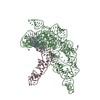
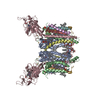
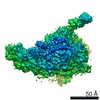
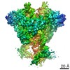
| Title | Cryo-EM structure of ENaC | ||||||||||||||||||||||||||||||||||||||||||||||||||||||||||||||||||||||||||||||||||||||||||
 Components Components |
| ||||||||||||||||||||||||||||||||||||||||||||||||||||||||||||||||||||||||||||||||||||||||||
 Keywords Keywords | MEMBRANE PROTEIN / sodium channel | ||||||||||||||||||||||||||||||||||||||||||||||||||||||||||||||||||||||||||||||||||||||||||
| Function / homology |  Function and homology information Function and homology informationsensory perception of salty taste / Sensory perception of salty taste / neutrophil-mediated killing of bacterium / aldosterone metabolic process / leukocyte activation involved in inflammatory response / cellular response to vasopressin / sensory perception of sour taste / cellular response to aldosterone / epithelial fluid transport / mucus secretion ...sensory perception of salty taste / Sensory perception of salty taste / neutrophil-mediated killing of bacterium / aldosterone metabolic process / leukocyte activation involved in inflammatory response / cellular response to vasopressin / sensory perception of sour taste / cellular response to aldosterone / epithelial fluid transport / mucus secretion / sodium channel complex / sodium ion homeostasis / renal system process / neutrophil activation involved in immune response / artery smooth muscle contraction / multicellular organismal-level water homeostasis / potassium ion homeostasis / intracellular sodium ion homeostasis / motile cilium / sodium ion import across plasma membrane / lung alveolus development / ligand-gated sodium channel activity / ciliary membrane / WW domain binding / erythrocyte homeostasis / response to food / ciliary transition zone / monoatomic ion channel activity / cellular response to acidic pH / cytoplasmic vesicle membrane / acrosomal vesicle / sodium ion transmembrane transport / regulation of blood pressure / multicellular organism growth / Stimuli-sensing channels / sperm principal piece / gene expression / apical plasma membrane / response to xenobiotic stimulus / external side of plasma membrane / extracellular exosome / nucleoplasm / plasma membrane / cytoplasm / cytosol Similarity search - Function | ||||||||||||||||||||||||||||||||||||||||||||||||||||||||||||||||||||||||||||||||||||||||||
| Biological species |  Homo sapiens (human) Homo sapiens (human) | ||||||||||||||||||||||||||||||||||||||||||||||||||||||||||||||||||||||||||||||||||||||||||
| Method | ELECTRON MICROSCOPY / single particle reconstruction / cryo EM / Resolution: 3.9 Å | ||||||||||||||||||||||||||||||||||||||||||||||||||||||||||||||||||||||||||||||||||||||||||
 Authors Authors | Noreng, S. / Bharadwaj, A. / Posert, R. / Yoshioka, C. / Baconguis, I. | ||||||||||||||||||||||||||||||||||||||||||||||||||||||||||||||||||||||||||||||||||||||||||
| Funding support |  United States, 1items United States, 1items
| ||||||||||||||||||||||||||||||||||||||||||||||||||||||||||||||||||||||||||||||||||||||||||
 Citation Citation | Journal: Elife / Year: 2018 Title: Structure of the human epithelial sodium channel by cryo-electron microscopy. Authors: Sigrid Noreng / Arpita Bharadwaj / Richard Posert / Craig Yoshioka / Isabelle Baconguis / Abstract: The epithelial sodium channel (ENaC), a member of the ENaC/DEG superfamily, regulates Na and water homeostasis. ENaCs assemble as heterotrimeric channels that harbor protease-sensitive domains ...The epithelial sodium channel (ENaC), a member of the ENaC/DEG superfamily, regulates Na and water homeostasis. ENaCs assemble as heterotrimeric channels that harbor protease-sensitive domains critical for gating the channel. Here, we present the structure of human ENaC in the uncleaved state determined by single-particle cryo-electron microscopy. The ion channel is composed of a large extracellular domain and a narrow transmembrane domain. The structure reveals that ENaC assembles with a 1:1:1 stoichiometry of α:β:γ subunits arranged in a counter-clockwise manner. The shape of each subunit is reminiscent of a hand with key gating domains of a 'finger' and a 'thumb.' Wedged between these domains is the elusive protease-sensitive inhibitory domain poised to regulate conformational changes of the 'finger' and 'thumb'; thus, the structure provides the first view of the architecture of inhibition of ENaC. | ||||||||||||||||||||||||||||||||||||||||||||||||||||||||||||||||||||||||||||||||||||||||||
| History |
|
- Structure visualization
| Movie |
Movie viewer |
|---|---|
| Structure viewer | Molecule: MolmilJmol/JSmol |
- Downloads & links
-Download
| PDBx/mmCIF format | 6bqn.cif.gz | 295.8 KB | Display | PDBx/mmCIF format |
|---|---|---|---|---|
| PDB format | pdb6bqn.ent.gz | 233.6 KB | Display | PDB format |
| PDBx/mmJSON format | 6bqn.json.gz | Tree view | PDBx/mmJSON format | |
| Others |  Other downloads Other downloads |
-Validation report
| Arichive directory | https://data.pdbj.org/pub/pdb/validation_reports/bq/6bqnftp://data.pdbj.org/pub/pdb/validation_reports/bq/6bqn | HTTPS FTP |
|---|
-Related structure data
| Related structure data | 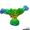 7130MC M: map data used to model this data C: citing same article ( |
|---|---|
| Similar structure data |


-Links
 PDBj
PDBj

- Assembly
| Deposited unit | 
|
|---|---|
| 1 |
|
-Components
-Antibody , 7 types, 7 molecules ABCDEFG
| #1: Antibody | Mass: 54038.953 Da / Num. of mol.: 1 Source method: isolated from a genetically manipulated source Details: Transmembrane helices have been built with poly UNK since the density for the transmembrane domain is not good enough to accurately build a model. Residues that have not been built is due to ...Details: Transmembrane helices have been built with poly UNK since the density for the transmembrane domain is not good enough to accurately build a model. Residues that have not been built is due to flexible areas of the protein where density is missing. The full sequence is described in sequence details. Source: (gene. exp.) Homo sapiens (human) / Production host: Homo sapiens (human) / References: UniProt: P37088*PLUS |
|---|---|
| #2: Antibody | Mass: 55038.633 Da / Num. of mol.: 1 Source method: isolated from a genetically manipulated source Details: Transmembrane helices have been built with poly UNK since the density for the transmembrane domain is not good enough to accurately build a model. Residues that have not been built is due to ...Details: Transmembrane helices have been built with poly UNK since the density for the transmembrane domain is not good enough to accurately build a model. Residues that have not been built is due to flexible areas of the protein where density is missing. The full sequence is described in sequence details. Source: (gene. exp.) Homo sapiens (human) / Production host: Homo sapiens (human) / References: UniProt: P51168*PLUS |
| #3: Antibody | Mass: 55834.457 Da / Num. of mol.: 1 Source method: isolated from a genetically manipulated source Details: The protein is EGFP-SCNN1G chimera. Transmembrane helices have been built with poly UNK since the density for the transmembrane domain is not good enough to accurately build a model. ...Details: The protein is EGFP-SCNN1G chimera. Transmembrane helices have been built with poly UNK since the density for the transmembrane domain is not good enough to accurately build a model. Residues that have not been built is due to flexible areas of the protein where density is missing. The first 260 residues is a His8 tag and a strep-tag followed by EGFP (UniProt accession code: C5MKY7). The full sequence is described in sequence details. Source: (gene. exp.) Homo sapiens (human) / Production host: Homo sapiens (human) / References: UniProt: P51170*PLUS |
| #4: Antibody | Mass: 9039.134 Da / Num. of mol.: 1 / Source method: isolated from a natural source / Details: Sequence unknown, built with poly UNK. / Source: (natural) |
| #5: Antibody | Mass: 10315.707 Da / Num. of mol.: 1 / Source method: isolated from a natural source / Details: Sequence unknown, built with poly UNK. / Source: (natural) |
| #6: Antibody | Mass: 9805.078 Da / Num. of mol.: 1 / Source method: isolated from a natural source / Details: Sequence unknown, built with poly UNK. / Source: (natural) |
| #7: Antibody | Mass: 9549.763 Da / Num. of mol.: 1 / Source method: isolated from a natural source / Details: Sequence unknown, built with poly UNK. / Source: (natural) |
-Details
| Has protein modification | Y |
|---|---|
| Sequence details | The sequence of SCNNIA is MAEEEALIEFHRSYRELFEFFANNTTIHGAIRLVSSQHNRMKTAFWAVLWLATFGMMYWQFGLLFGEY ...The sequence of SCNNIA is MAEEEALIEF |
-Experimental details
-Experiment
| Experiment | Method: ELECTRON MICROSCOPY |
|---|---|
| EM experiment | Aggregation state: PARTICLE / 3D reconstruction method: single particle reconstruction |
- Sample preparation
| Component | Name: Membrane protein complex / Type: CELL / Entity ID: all / Source: RECOMBINANT |
|---|---|
| Source (natural) | Organism: Homo sapiens (human) |
| Source (recombinant) | Organism: Homo sapiens (human) |
| Buffer solution | pH: 8 |
| Specimen | Embedding applied: NO / Shadowing applied: NO / Staining applied: NO / Vitrification applied: YES |
| Vitrification | Cryogen name: ETHANE |
- Electron microscopy imaging
| Experimental equipment |  Model: Titan Krios / Image courtesy: FEI Company |
|---|---|
| Microscopy | Model: FEI TITAN KRIOS |
| Electron gun | Electron source:  FIELD EMISSION GUN / Accelerating voltage: 300 kV / Illumination mode: OTHER FIELD EMISSION GUN / Accelerating voltage: 300 kV / Illumination mode: OTHER |
| Electron lens | Mode: OTHER |
| Image recording | Electron dose: 60 e/Å2 / Film or detector model: GATAN K2 SUMMIT (4k x 4k) |
- Processing
| Software | Name: PHENIX / Version: 1.13_2998: / Classification: refinement | ||||||||||||||||||||||||
|---|---|---|---|---|---|---|---|---|---|---|---|---|---|---|---|---|---|---|---|---|---|---|---|---|---|
| EM software | Name: PHENIX / Category: model refinement | ||||||||||||||||||||||||
| CTF correction | Type: PHASE FLIPPING AND AMPLITUDE CORRECTION | ||||||||||||||||||||||||
| 3D reconstruction | Resolution: 3.9 Å / Resolution method: FSC 0.143 CUT-OFF / Num. of particles: 304615 / Symmetry type: POINT | ||||||||||||||||||||||||
| Refine LS restraints |
|
