 Movie
Movie Controller


+ Open data

- Basic information
| Entry | Database: PDB / ID: 6bdu | ||||||
|---|---|---|---|---|---|---|---|
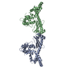
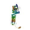
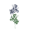
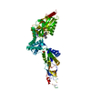
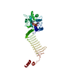
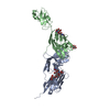
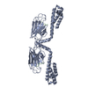
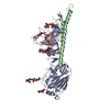
| Title | Crystal structure of PprA from Deinococcus radiodurans | ||||||
 Components Components | DNA repair protein PprA | ||||||
 Keywords Keywords | DNA BINDING PROTEIN / DNA damage repair / Radiation induced / Genome segregation / Filment formation | ||||||
| Function / homology | cellular response to desiccation / cellular response to gamma radiation / double-strand break repair via nonhomologous end joining / double-stranded DNA binding / damaged DNA binding / DNA repair / DNA repair protein PprA Function and homology information Function and homology information | ||||||
| Biological species |  Deinococcus radiodurans (radioresistant) Deinococcus radiodurans (radioresistant) | ||||||
| Method |  X-RAY DIFFRACTION / SYNCHROTRON / SAD / Resolution: 2 Å X-RAY DIFFRACTION / SYNCHROTRON / SAD / Resolution: 2 Å | ||||||
 Authors Authors | Szabla, R. / Czerwinski, M. / Junop, M.S. | ||||||
| Funding support |  Canada, 1items Canada, 1items
| ||||||
 Citation Citation | Journal: To Be Published Title: Crystal structure of PprA from Deinococcus radiodurans Authors: Szabla, R. / Czerwinski, M. / Junop, M.S. | ||||||
| History |
|
- Structure visualization
| Structure viewer | Molecule: MolmilJmol/JSmol |
|---|
- Downloads & links
-Download
| PDBx/mmCIF format | 6bdu.cif.gz | 124.6 KB | Display | PDBx/mmCIF format |
|---|---|---|---|---|
| PDB format | pdb6bdu.ent.gz | 95.7 KB | Display | PDB format |
| PDBx/mmJSON format | 6bdu.json.gz | Tree view | PDBx/mmJSON format | |
| Others |  Other downloads Other downloads |
-Validation report
| Summary document | 6bdu_validation.pdf.gz | 436.2 KB | Display | wwPDB validaton report |
|---|---|---|---|---|
| Full document | 6bdu_full_validation.pdf.gz | 439.7 KB | Display | |
| Data in XML | 6bdu_validation.xml.gz | 24 KB | Display | |
| Data in CIF | 6bdu_validation.cif.gz | 34.9 KB | Display | |
| Arichive directory | https://data.pdbj.org/pub/pdb/validation_reports/bd/6bduftp://data.pdbj.org/pub/pdb/validation_reports/bd/6bdu | HTTPS FTP |
-Related structure data


-Links
 PDBj
PDBj- Assembly
| Deposited unit | 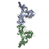
| ||||||||
|---|---|---|---|---|---|---|---|---|---|
| 1 |
| ||||||||
| Unit cell |
|
-Components
| #1: Protein | Mass: 33602.750 Da / Num. of mol.: 2 / Mutation: D180K, D184K Source method: isolated from a genetically manipulated source Source: (gene. exp.) Deinococcus radiodurans (radioresistant)Strain: ATCC 13939 / DSM 20539 / JCM 16871 / LMG 4051 / NBRC 15346 / NCIMB 9279 / R1 / VKM B-1422 Gene: pprA, DR_A0346 / Plasmid: pDEST-527 / Cell line (production host): B834(DE3) / Production host: #2: Water | ChemComp-HOH / |  Mass: 18.015 Da / Num. of mol.: 376 / Source method: isolated from a natural source / Formula: H2O Mass: 18.015 Da / Num. of mol.: 376 / Source method: isolated from a natural source / Formula: H2OHas protein modification | Y | |
|---|
-Experimental details
-Experiment
| Experiment | Method: X-RAY DIFFRACTION / Number of used crystals: 1 |
|---|
- Sample preparation
| Crystal | Density Matthews: 2.44 Å3/Da / Density % sol: 49.53 % / Description: square prism |
|---|---|
| Crystal grow | Temperature: 293.15 K / Method: vapor diffusion, hanging drop / pH: 8 Details: Protein at 2.4mg/mL in 150mM KCl, 20mM Tris, pH 7.5 was mixed in 1:1 volume ratio with a solution of 0.2 M Lithium Citrate Tribasic and 20 % (w/v) PEG 3350. The drop was suspended over 1.5M Ammonium sulfate. |
-Data collection
| Diffraction | Mean temperature: 100 K |
|---|---|
| Diffraction source | Source: SYNCHROTRON / Site: APS  / Beamline: 17-ID / Wavelength: 0.9762 Å / Beamline: 17-ID / Wavelength: 0.9762 Å |
| Detector | Type: DECTRIS PILATUS 6M / Detector: PIXEL / Date: Apr 22, 2017 |
| Radiation | Protocol: SINGLE WAVELENGTH / Monochromatic (M) / Laue (L): M / Scattering type: x-ray |
| Radiation wavelength | Wavelength: 0.9762 Å / Relative weight: 1 |
| Reflection | Resolution: 2→58.34 Å / Num. obs: 45262 / % possible obs: 100 % / Redundancy: 12.1 % / CC1/2: 0.997 / Rmerge(I) obs: 0.152 / Rpim(I) all: 0.061 / Rrim(I) all: 0.159 / Net I/σ(I): 11.2 |
| Reflection shell | Resolution: 2→2.05 Å / Redundancy: 12.6 % / Rmerge(I) obs: 1.628 / Mean I/σ(I) obs: 1.9 / Num. unique obs: 3281 / CC1/2: 0.658 / Rpim(I) all: 0.662 / Rrim(I) all: 1.606 |
- Processing
| Software |
| ||||||||||||||||||||||||||||||||||||||||||||||||||||||||||||||||||||||||||||||||||||||||||||||||||||||||||||||||||||||||||||||
|---|---|---|---|---|---|---|---|---|---|---|---|---|---|---|---|---|---|---|---|---|---|---|---|---|---|---|---|---|---|---|---|---|---|---|---|---|---|---|---|---|---|---|---|---|---|---|---|---|---|---|---|---|---|---|---|---|---|---|---|---|---|---|---|---|---|---|---|---|---|---|---|---|---|---|---|---|---|---|---|---|---|---|---|---|---|---|---|---|---|---|---|---|---|---|---|---|---|---|---|---|---|---|---|---|---|---|---|---|---|---|---|---|---|---|---|---|---|---|---|---|---|---|---|---|---|---|---|
| Refinement | Method to determine structure: SAD / Resolution: 2→36.21 Å / SU ML: 0.26 / Cross valid method: FREE R-VALUE / σ(F): 1.34 / Phase error: 24.34
| ||||||||||||||||||||||||||||||||||||||||||||||||||||||||||||||||||||||||||||||||||||||||||||||||||||||||||||||||||||||||||||||
| Solvent computation | Shrinkage radii: 0.9 Å / VDW probe radii: 1.11 Å | ||||||||||||||||||||||||||||||||||||||||||||||||||||||||||||||||||||||||||||||||||||||||||||||||||||||||||||||||||||||||||||||
| Refinement step | Cycle: LAST / Resolution: 2→36.21 Å
| ||||||||||||||||||||||||||||||||||||||||||||||||||||||||||||||||||||||||||||||||||||||||||||||||||||||||||||||||||||||||||||||
| Refine LS restraints |
| ||||||||||||||||||||||||||||||||||||||||||||||||||||||||||||||||||||||||||||||||||||||||||||||||||||||||||||||||||||||||||||||
| LS refinement shell |
|
