 Movie
Movie Controller


+ Open data

- Basic information
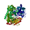
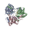
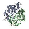
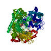
| Entry | Database: PDB / ID: 6b9l | |||||||||
|---|---|---|---|---|---|---|---|---|---|---|
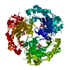
| Title | Crystal structure of EphA2 with peptide 135E2 | |||||||||
 Components Components |
| |||||||||
 Keywords Keywords | TRANSFERASE / complex / ligand binding domain | |||||||||
| Function / homology |  Function and homology information Function and homology informationnotochord cell development / notochord formation / blood vessel endothelial cell proliferation involved in sprouting angiogenesis / negative regulation of lymphangiogenesis / lens fiber cell morphogenesis / axial mesoderm formation / cAMP metabolic process / regulation of blood vessel endothelial cell migration / pericyte cell differentiation / leading edge membrane ...notochord cell development / notochord formation / blood vessel endothelial cell proliferation involved in sprouting angiogenesis / negative regulation of lymphangiogenesis / lens fiber cell morphogenesis / axial mesoderm formation / cAMP metabolic process / regulation of blood vessel endothelial cell migration / pericyte cell differentiation / leading edge membrane / negative regulation of chemokine production / ephrin receptor activity / activation of GTPase activity / response to growth factor / post-anal tail morphogenesis / bone remodeling / positive regulation of bicellular tight junction assembly / regulation of lamellipodium assembly / negative regulation of cell adhesion mediated by integrin / branching involved in mammary gland duct morphogenesis / EPH-Ephrin signaling / central nervous system neuron differentiation / RND1 GTPase cycle / RND2 GTPase cycle / RND3 GTPase cycle / neural tube development / mammary gland epithelial cell proliferation / tight junction / RHOV GTPase cycle / EPHA-mediated growth cone collapse / growth factor binding / RHOU GTPase cycle / lamellipodium membrane / RHOG GTPase cycle / EPH-ephrin mediated repulsion of cells / RAC3 GTPase cycle / regulation of angiogenesis / RAC2 GTPase cycle / ephrin receptor signaling pathway / vasculogenesis / regulation of ERK1 and ERK2 cascade / keratinocyte differentiation / RAC1 GTPase cycle / transmembrane receptor protein tyrosine kinase activity / osteoclast differentiation / cell surface receptor protein tyrosine kinase signaling pathway / negative regulation of angiogenesis / molecular function activator activity / protein localization to plasma membrane / positive regulation of protein localization to plasma membrane / cell chemotaxis / skeletal system development / cell motility / receptor protein-tyrosine kinase / intrinsic apoptotic signaling pathway in response to DNA damage / ruffle membrane / osteoblast differentiation / cell migration / lamellipodium / virus receptor activity / angiogenesis / cell adhesion / signaling receptor complex / defense response to Gram-positive bacterium / positive regulation of cell migration / cadherin binding / inflammatory response / focal adhesion / cell surface / ATP binding / plasma membrane Similarity search - Function | |||||||||
| Biological species |  Homo sapiens (human) Homo sapiens (human)unidentified (others) | |||||||||
| Method |  X-RAY DIFFRACTION / SYNCHROTRON / Resolution: 3.2 Å X-RAY DIFFRACTION / SYNCHROTRON / Resolution: 3.2 Å | |||||||||
 Authors Authors | Song, J. / Tan, X. | |||||||||
 Citation Citation | Journal: ACS Chem. Biol. / Year: 2018 Title: Structure-Based Design of Novel EphA2 Agonistic Agents with Nanomolar Affinity in Vitro and in Cell. Authors: Gambini, L. / Salem, A.F. / Udompholkul, P. / Tan, X.F. / Baggio, C. / Shah, N. / Aronson, A. / Song, J. / Pellecchia, M. | |||||||||
| History |
|
- Structure visualization
| Structure viewer | Molecule: MolmilJmol/JSmol |
|---|
- Downloads & links
-Download
| PDBx/mmCIF format | 6b9l.cif.gz | 305.3 KB | Display | PDBx/mmCIF format |
|---|---|---|---|---|
| PDB format | pdb6b9l.ent.gz | 251.4 KB | Display | PDB format |
| PDBx/mmJSON format | 6b9l.json.gz | Tree view | PDBx/mmJSON format | |
| Others |  Other downloads Other downloads |
-Validation report
| Arichive directory | https://data.pdbj.org/pub/pdb/validation_reports/b9/6b9lftp://data.pdbj.org/pub/pdb/validation_reports/b9/6b9l | HTTPS FTP |
|---|
-Related structure data
| Similar structure data |
|---|


-Links
 PDBj
PDBj


- Assembly
| Deposited unit | 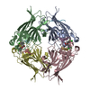
| ||||||||
|---|---|---|---|---|---|---|---|---|---|
| 1 |
| ||||||||
| Unit cell |
|
-Components
| #1: Protein | Mass: 22242.193 Da / Num. of mol.: 4 Source method: isolated from a genetically manipulated source Source: (gene. exp.) Homo sapiens (human) / Gene: EPHA2, ECKProduction host:  References: UniProt: P29317, receptor protein-tyrosine kinase #2: Protein/peptide | Mass: 1325.806 Da / Num. of mol.: 4 Source method: isolated from a genetically manipulated source Source: (gene. exp.) unidentified (others) / Production host: #3: Protein/peptide | | Mass: 712.758 Da / Num. of mol.: 1 Source method: isolated from a genetically manipulated source Source: (gene. exp.) Homo sapiens (human)Production host: Has protein modification | Y | |
|---|
-Experimental details
-Experiment
| Experiment | Method: X-RAY DIFFRACTION / Number of used crystals: 1 |
|---|
- Sample preparation
| Crystal | Density Matthews: 2.98 Å3/Da / Density % sol: 62.79 % |
|---|---|
| Crystal grow | Temperature: 277 K / Method: vapor diffusion, hanging drop / pH: 8 / Details: Lithium sulphate, Tris-Cl |
-Data collection
| Diffraction | Mean temperature: 100 K |
|---|---|
| Diffraction source | Source: SYNCHROTRON / Site: ALS  / Beamline: 5.0.3 / Wavelength: 0.97741 Å / Beamline: 5.0.3 / Wavelength: 0.97741 Å |
| Detector | Type: ADSC QUANTUM 315r / Detector: CCD / Date: Aug 15, 2017 |
| Radiation | Protocol: SINGLE WAVELENGTH / Monochromatic (M) / Laue (L): M / Scattering type: x-ray |
| Radiation wavelength | Wavelength: 0.97741 Å / Relative weight: 1 |
| Reflection | Resolution: 3.2→50 Å / Num. obs: 18465 / % possible obs: 96 % / Redundancy: 3.5 % / Net I/σ(I): 3.9 |
- Processing
| Software |
| ||||||||||||||||||||||||||||||||||||||||||||||||||||||||||||||||||||||||||||||||||||||||||||||||||
|---|---|---|---|---|---|---|---|---|---|---|---|---|---|---|---|---|---|---|---|---|---|---|---|---|---|---|---|---|---|---|---|---|---|---|---|---|---|---|---|---|---|---|---|---|---|---|---|---|---|---|---|---|---|---|---|---|---|---|---|---|---|---|---|---|---|---|---|---|---|---|---|---|---|---|---|---|---|---|---|---|---|---|---|---|---|---|---|---|---|---|---|---|---|---|---|---|---|---|---|
| Refinement | Resolution: 3.2→47.08 Å / SU ML: 0.44 / Cross valid method: FREE R-VALUE / σ(F): 1.35 / Phase error: 26.16 / Stereochemistry target values: ML
| ||||||||||||||||||||||||||||||||||||||||||||||||||||||||||||||||||||||||||||||||||||||||||||||||||
| Solvent computation | Shrinkage radii: 0.9 Å / VDW probe radii: 1.11 Å / Solvent model: FLAT BULK SOLVENT MODEL | ||||||||||||||||||||||||||||||||||||||||||||||||||||||||||||||||||||||||||||||||||||||||||||||||||
| Refinement step | Cycle: LAST / Resolution: 3.2→47.08 Å
| ||||||||||||||||||||||||||||||||||||||||||||||||||||||||||||||||||||||||||||||||||||||||||||||||||
| Refine LS restraints |
| ||||||||||||||||||||||||||||||||||||||||||||||||||||||||||||||||||||||||||||||||||||||||||||||||||
| LS refinement shell |
|
