| 登録情報 | データベース: PDB / ID: 6b1z
|
|---|
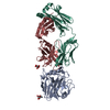
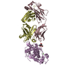
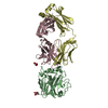
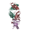
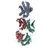
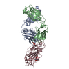
| タイトル | Crystal Structure of Glutamate-tRNA Synthetase from Elizabethkingia anophelis |
|---|
 要素 要素 | Glutamate--tRNA ligase |
|---|
 キーワード キーワード | LIGASE / SSGCID / Elizabethkingia anophelis / Glutamate--tRNA ligase / Structural Genomics / Seattle Structural Genomics Center for Infectious Disease |
|---|
| 機能・相同性 |  機能・相同性情報 機能・相同性情報
glutamate-tRNA ligase / glutamate-tRNA ligase activity / glutamyl-tRNA aminoacylation / tRNA binding / zinc ion binding / ATP binding / cytoplasm類似検索 - 分子機能 Glutamate-tRNA ligase, bacterial/mitochondrial / Glutamyl-tRNA synthetase / Aminoacyl-tRNA synthetase, class I, anticodon-binding superfamily / Aminoacyl-tRNA synthetase, class I, anticodon-binding domain, subdomain 2 / Aminoacyl-tRNA synthetase, class I, anticodon-binding / Anticodon binding domain / : / Glutamine-tRNA ligase, alpha-bundle domain superfamily / Glutamyl-tRNA Synthetase; domain 2 / Glutamyl-trna Synthetase; Domain 2 ...Glutamate-tRNA ligase, bacterial/mitochondrial / Glutamyl-tRNA synthetase / Aminoacyl-tRNA synthetase, class I, anticodon-binding superfamily / Aminoacyl-tRNA synthetase, class I, anticodon-binding domain, subdomain 2 / Aminoacyl-tRNA synthetase, class I, anticodon-binding / Anticodon binding domain / : / Glutamine-tRNA ligase, alpha-bundle domain superfamily / Glutamyl-tRNA Synthetase; domain 2 / Glutamyl-trna Synthetase; Domain 2 / Glutamyl-tRNA Synthetase; domain 3 / Glutamyl-tRNA Synthetase; Domain 3 / Glutamyl/glutaminyl-tRNA synthetase / Glutamyl/glutaminyl-tRNA synthetase, class Ib, catalytic domain / tRNA synthetases class I (E and Q), catalytic domain / Aminoacyl-tRNA synthetase, class I, conserved site / Aminoacyl-transfer RNA synthetases class-I signature. / HUPs / Rossmann-like alpha/beta/alpha sandwich fold / Alpha-Beta Complex / Rossmann fold / Orthogonal Bundle / 3-Layer(aba) Sandwich / Mainly Alpha / Alpha Beta類似検索 - ドメイン・相同性 FORMIC ACID / Glutamate--tRNA ligase / Glutamate--tRNA ligase類似検索 - 構成要素 |
|---|
| 生物種 |  Elizabethkingia anophelis (バクテリア) Elizabethkingia anophelis (バクテリア) |
|---|
| 手法 |  X線回折 / シンクロトロン / 分子置換 / 解像度: 1.6 Å X線回折 / シンクロトロン / 分子置換 / 解像度: 1.6 Å |
|---|
 データ登録者 データ登録者 | Seattle Structural Genomics Center for Infectious Disease (SSGCID) |
|---|
 引用 引用 | ジャーナル: Acta Crystallogr.,Sect.F / 年: 2022
タイトル: Crystal structures of glutamyl-tRNA synthetase from Elizabethkingia anopheles and E. meningosepticum.
著者: Brooks, L. / Subramanian, S. / Dranow, D.M. / Mayclin, S.J. / Myler, P.J. / Asojo, O.A. |
|---|
| 履歴 | | 登録 | 2017年9月19日 | 登録サイト: RCSB / 処理サイト: RCSB |
|---|
| 改定 1.0 | 2017年10月4日 | Provider: repository / タイプ: Initial release |
|---|
| 改定 1.1 | 2018年4月4日 | Group: Data collection / カテゴリ: diffrn_source
Item: _diffrn_source.pdbx_synchrotron_beamline / _diffrn_source.type |
|---|
| 改定 1.2 | 2023年10月4日 | Group: Data collection / Database references ...Data collection / Database references / Derived calculations / Refinement description
カテゴリ: chem_comp_atom / chem_comp_bond ...chem_comp_atom / chem_comp_bond / database_2 / pdbx_initial_refinement_model / pdbx_struct_conn_angle / struct_conn
Item: _database_2.pdbx_DOI / _database_2.pdbx_database_accession ..._database_2.pdbx_DOI / _database_2.pdbx_database_accession / _pdbx_struct_conn_angle.ptnr1_auth_seq_id / _pdbx_struct_conn_angle.ptnr1_symmetry / _pdbx_struct_conn_angle.ptnr3_auth_seq_id / _pdbx_struct_conn_angle.ptnr3_symmetry / _pdbx_struct_conn_angle.value / _struct_conn.pdbx_dist_value / _struct_conn.ptnr2_auth_seq_id / _struct_conn.ptnr2_symmetry |
|---|
| 改定 1.3 | 2024年7月3日 | Group: Database references / カテゴリ: citation / citation_author
Item: _citation.country / _citation.journal_abbrev ..._citation.country / _citation.journal_abbrev / _citation.journal_id_ASTM / _citation.journal_id_CSD / _citation.journal_id_ISSN / _citation.journal_volume / _citation.page_first / _citation.page_last / _citation.pdbx_database_id_DOI / _citation.pdbx_database_id_PubMed / _citation.title / _citation.year |
|---|
|
|---|
 ムービー
ムービー コントローラー


 データを開く
 基本情報
基本情報 構造の表示
構造の表示 ダウンロードとリンク
 その他のダウンロード


 PDBj
PDBj
 集合体
集合体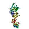

 分子量: 46.025 Da / 分子数: 16 / 由来タイプ: 合成 / 式: CH2O2
 分子量: 62.068 Da / 分子数: 7 / 由来タイプ: 合成 / 式: C2H6O2
 分子量: 24.305 Da / 分子数: 1 / 由来タイプ: 合成 / 式: Mg
 分子量: 18.015 Da / 分子数: 576 / 由来タイプ: 天然 / 式: H2O
 試料調製
試料調製 / ビームライン: 21-ID-D / 波長: 0.97872 Å
 解析
解析