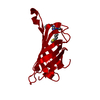
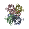
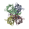
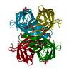
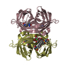
[English] 日本語
Yorodumi
- PDB-6anx: Peroxide Activation Regulated by Hydrogen Bonds within Artificial... -

+
Open data


ID or keywords:

Loading...

-
Basic information

Entry
Database: PDB / ID: 6anx
TitlePeroxide Activation Regulated by Hydrogen Bonds within Artificial Cu Proteins - WT (low exposure)
ComponentsStreptavidin
KeywordsMETAL BINDING PROTEIN / streptavidin / biotin / copper / hydroperoxo / secondary coordination sphere / hydrogen bond / biotin binding protein
Function / homology
Function and homology information


biotin binding / extracellular region
Similarity search - Function
Avidin-like / Avidin-like, conserved site / Avidin-like domain signature. / Avidin / Avidin/streptavidin / Avidin-like superfamily / Avidin family / Avidin-like domain profile. / Lipocalin / Beta Barrel / Mainly Beta
Similarity search - Domain/homology
ACETATE ION / Chem-SI0 / Streptavidin
Similarity search - Component
Biological speciesStreptomyces avidinii (bacteria)
MethodX-RAY DIFFRACTION / SYNCHROTRON / MOLECULAR REPLACEMENT / Resolution: 1.62 Å
AuthorsMann, S.I. / Heinisch, T. / Ward, T.R. / Borovik, A.S.
Funding support United States, 1items
OrganizationGrant numberCountry
National Institutes of Health/National Institute of General Medical Sciences (NIH/NIGMS)GM-120349 United States
CitationJournal: J. Am. Chem. Soc. / Year: 2017
Title: Peroxide Activation Regulated by Hydrogen Bonds within Artificial Cu Proteins.
Authors: Mann, S.I. / Heinisch, T. / Ward, T.R. / Borovik, A.S.
History
DepositionAug 14, 2017Deposition site: RCSB / Processing site: RCSB
Revision 1.0Nov 22, 2017Provider: repository / Type: Initial release
Revision 1.1Dec 13, 2017Group: Database references / Category: citation
Item: _citation.journal_volume / _citation.page_first / _citation.page_last
Revision 1.2Jan 1, 2020Group: Author supporting evidence / Category: pdbx_audit_support / Item: _pdbx_audit_support.funding_organization
Revision 1.3Oct 4, 2023Group: Data collection / Database references / Refinement description
Category: chem_comp_atom / chem_comp_bond ...chem_comp_atom / chem_comp_bond / database_2 / pdbx_initial_refinement_model
Item: _database_2.pdbx_DOI / _database_2.pdbx_database_accession

-
Structure visualization

Structure viewerMolecule:
MolmilJmol/JSmol

Downloads & links

-
Assembly

Deposited unit
A: Streptavidin
hetero molecules


Theoretical massNumber of molelcules
Total (without water)17,3344
Polymers16,5701
Non-polymers7643
Water1,78399
1
A: Streptavidin
hetero molecules

A: Streptavidin
hetero molecules

A: Streptavidin
hetero molecules

A: Streptavidin
hetero molecules


Theoretical massNumber of molelcules
Total (without water)69,33716
Polymers66,2804
Non-polymers3,05712
Water724
TypeNameSymmetry operationNumber
identity operation1_555x,y,z1
crystal symmetry operation8_665-y+1,-x+1,-z1
crystal symmetry operation10_665-x+1,-y+1,z1
crystal symmetry operation15_555y,x,-z1
Buried area9390 Å2
ΔGint-55 kcal/mol
Surface area19410 Å2
MethodPISA
Unit cell
Length a, b, c (Å)57.640, 57.640, 183.610
Angle α, β, γ (deg.)90.00, 90.00, 90.00
Int Tables number98
Space group name H-MI4122
Components on special symmetry positions
IDModelComponents
11A-332-

HOH

-
Components

#1: Protein Streptavidin /


Mass: 16570.018 Da / Num. of mol.: 1 / Fragment: UNP residues 38-183
Source method: isolated from a genetically manipulated source
Source: (gene. exp.) Streptomyces avidinii (bacteria) / Production host: Escherichia coli (E. coli) / References: UniProt: P22629
#2: Chemical ChemComp-SI0 / [N-(2-{bis[2-(pyridin-2-yl-kappaN)ethyl]amino-kappaN}ethyl)-5-(2-oxohexahydro-1H-thieno[3,4-d]imidazol-4-yl)pentanamide](hydrogen peroxido-kappaO)hydroxycopper


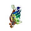
Mass: 609.220 Da / Num. of mol.: 1 / Source method: obtained synthetically / Formula: C26H37CuN6O5S / Feature type: SUBJECT OF INVESTIGATION
#3: Chemical ChemComp-ACT / ACETATE ION / Acetate


Mass: 59.044 Da / Num. of mol.: 1 / Source method: obtained synthetically / Formula: C2H3O2
#4: Chemical ChemComp-SO4 / SULFATE ION / Sulfate


Mass: 96.063 Da / Num. of mol.: 1 / Source method: obtained synthetically / Formula: SO4
#5: Water ChemComp-HOH / water / Water


Mass: 18.015 Da / Num. of mol.: 99 / Source method: isolated from a natural source / Formula: H2O

-
Experimental details

-
Experiment

ExperimentMethod: X-RAY DIFFRACTION / Number of used crystals: 1

-
Sample preparation

CrystalDensity Matthews: 2.3 Å3/Da / Density % sol: 46.54 %
Crystal growTemperature: 274 K / Method: vapor diffusion, sitting drop / pH: 4 / Details: 2.6 M ammonium sulfate, 0.1 M sodium acetate

-
Data collection

DiffractionMean temperature: 100 K
Diffraction sourceSource: SYNCHROTRON / Site: ALS / Beamline: 8.2.2 / Wavelength: 1 Å
DetectorType: ADSC QUANTUM 315r / Detector: CCD / Date: Aug 11, 2017
RadiationMonochromator: double crystal Si(111) / Protocol: SINGLE WAVELENGTH / Monochromatic (M) / Laue (L): M / Scattering type: x-ray
Radiation wavelengthWavelength: 1 Å / Relative weight: 1
ReflectionResolution: 1.62→54.99 Å / Num. obs: 20471 / % possible obs: 100 % / Redundancy: 13.4 % / Net I/σ(I): 12.9
Reflection shellResolution: 1.62→1.64 Å / Redundancy: 10.1 % / Mean I/σ(I) obs: 1.1 / Num. unique obs: 995 / % possible all: 99.5

-
Processing

Software
NameVersionClassification
REFMAC5.8.0135refinement
iMOSFLMdata reduction
Aimlessdata scaling
PHASERphasing
RefinementMethod to determine structure: MOLECULAR REPLACEMENT
Starting model: PDB entry 2QCB
Resolution: 1.62→54.99 Å / Cor.coef. Fo:Fc: 0.969 / Cor.coef. Fo:Fc free: 0.959 / SU B: 1.617 / SU ML: 0.052 / Cross valid method: THROUGHOUT / ESU R: 0.072 / ESU R Free: 0.073 / Details: HYDROGENS HAVE BEEN ADDED IN THE RIDING POSITIONS
RfactorNum. reflection% reflectionSelection details
Rfree0.18691 1057 5.2 %RANDOM
Rwork0.16318 ---
obs0.16439 19386 99.95 %-
Solvent computationIon probe radii: 0.8 Å / Shrinkage radii: 0.8 Å / VDW probe radii: 1.2 Å
Displacement parametersBiso mean: 22.085 Å2
Baniso -1Baniso -2Baniso -3
1--0 Å20 Å2-0 Å2
2---0 Å2-0 Å2
3---0.01 Å2
Refinement stepCycle: 1 / Resolution: 1.62→54.99 Å
ProteinNucleic acidLigandSolventTotal
Num. atoms936 0 48 99 1083
Refine LS restraints
Refine-IDTypeDev idealDev ideal targetNumber
X-RAY DIFFRACTIONr_bond_refined_d0.0230.021024
X-RAY DIFFRACTIONr_bond_other_d0.0020.02863
X-RAY DIFFRACTIONr_angle_refined_deg2.3442.0471405
X-RAY DIFFRACTIONr_angle_other_deg1.10931972
X-RAY DIFFRACTIONr_dihedral_angle_1_deg8.0965128
X-RAY DIFFRACTIONr_dihedral_angle_2_deg30.36723.57142
X-RAY DIFFRACTIONr_dihedral_angle_3_deg13.70415135
X-RAY DIFFRACTIONr_dihedral_angle_4_deg17.085155
X-RAY DIFFRACTIONr_chiral_restr0.130.2148
X-RAY DIFFRACTIONr_gen_planes_refined0.0120.021169
X-RAY DIFFRACTIONr_gen_planes_other0.0020.02248
X-RAY DIFFRACTIONr_nbd_refined
X-RAY DIFFRACTIONr_nbd_other
X-RAY DIFFRACTIONr_nbtor_refined
X-RAY DIFFRACTIONr_nbtor_other
X-RAY DIFFRACTIONr_xyhbond_nbd_refined
X-RAY DIFFRACTIONr_xyhbond_nbd_other
X-RAY DIFFRACTIONr_metal_ion_refined
X-RAY DIFFRACTIONr_metal_ion_other
X-RAY DIFFRACTIONr_symmetry_vdw_refined
X-RAY DIFFRACTIONr_symmetry_vdw_other
X-RAY DIFFRACTIONr_symmetry_hbond_refined
X-RAY DIFFRACTIONr_symmetry_hbond_other
X-RAY DIFFRACTIONr_symmetry_metal_ion_refined
X-RAY DIFFRACTIONr_symmetry_metal_ion_other
X-RAY DIFFRACTIONr_mcbond_it2.6091.916504
X-RAY DIFFRACTIONr_mcbond_other2.5131.903502
X-RAY DIFFRACTIONr_mcangle_it3.962.84628
X-RAY DIFFRACTIONr_mcangle_other3.9582.849629
X-RAY DIFFRACTIONr_scbond_it3.8042.302520
X-RAY DIFFRACTIONr_scbond_other3.7732.282517
X-RAY DIFFRACTIONr_scangle_it
X-RAY DIFFRACTIONr_scangle_other5.4683.254769
X-RAY DIFFRACTIONr_long_range_B_refined7.46217.0761208
X-RAY DIFFRACTIONr_long_range_B_other7.45917.11209
X-RAY DIFFRACTIONr_rigid_bond_restr
X-RAY DIFFRACTIONr_sphericity_free
X-RAY DIFFRACTIONr_sphericity_bonded
LS refinement shellResolution: 1.615→1.657 Å / Total num. of bins used: 20
RfactorNum. reflection% reflection
Rfree0.366 73 -
Rwork0.311 1400 -
obs--99.33 %

+
About Yorodumi

-
News

-
Feb 9, 2022. New format data for meta-information of EMDB entries

New format data for meta-information of EMDB entries

  • Version 3 of the EMDB header file is now the official format.
  • The previous official version 1.9 will be removed from the archive.

Related info.:EMDB header

External links:wwPDB to switch to version 3 of the EMDB data model

-
Aug 12, 2020. Covid-19 info

Covid-19 info

URL: https://pdbj.org/emnavi/covid19.php

New page: Covid-19 featured information page in EM Navigator.

Related info.:Covid-19 info / Mar 5, 2020. Novel coronavirus structure data

+
Mar 5, 2020. Novel coronavirus structure data

Novel coronavirus structure data

Related info.:Yorodumi Speices / Aug 12, 2020. Covid-19 info

External links:COVID-19 featured content - PDBj / Molecule of the Month (242):Coronavirus Proteases

+
Jan 31, 2019. EMDB accession codes are about to change! (news from PDBe EMDB page)

EMDB accession codes are about to change! (news from PDBe EMDB page)

  • The allocation of 4 digits for EMDB accession codes will soon come to an end. Whilst these codes will remain in use, new EMDB accession codes will include an additional digit and will expand incrementally as the available range of codes is exhausted. The current 4-digit format prefixed with “EMD-” (i.e. EMD-XXXX) will advance to a 5-digit format (i.e. EMD-XXXXX), and so on. It is currently estimated that the 4-digit codes will be depleted around Spring 2019, at which point the 5-digit format will come into force.
  • The EM Navigator/Yorodumi systems omit the EMD- prefix.

Related info.:Q: What is EMD? / ID/Accession-code notation in Yorodumi/EM Navigator

External links:EMDB Accession Codes are Changing Soon! / Contact to PDBj

+
Jul 12, 2017. Major update of PDB

Major update of PDB

  • wwPDB released updated PDB data conforming to the new PDBx/mmCIF dictionary.
  • This is a major update changing the version number from 4 to 5, and with Remediation, in which all the entries are updated.
  • In this update, many items about electron microscopy experimental information are reorganized (e.g. em_software).
  • Now, EM Navigator and Yorodumi are based on the updated data.

External links:wwPDB Remediation / Enriched Model Files Conforming to OneDep Data Standards Now Available in the PDB FTP Archive

-
Yorodumi

Thousand views of thousand structures

  • Yorodumi is a browser for structure data from EMDB, PDB, SASBDB, etc.
  • This page is also the successor to EM Navigator detail page, and also detail information page/front-end page for Omokage search.
  • The word "yorodu" (or yorozu) is an old Japanese word meaning "ten thousand". "mi" (miru) is to see.

Related info.:EMDB / PDB / SASBDB / Comparison of 3 databanks / Yorodumi Search / Aug 31, 2016. New EM Navigator & Yorodumi / Yorodumi Papers / Jmol/JSmol / Function and homology information / Changes in new EM Navigator and Yorodumi

Read more