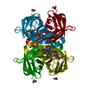
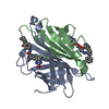
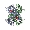
Entry Database : PDB / ID : 5vl8Title Coordination Chemistry within a Protein Host: Regulation of the Secondary Coordination Sphere Streptavidin Keywords / / / / / / Function / homology Function Domain/homology Component
/ / / / / / / / / / / / / / / / / / / / Biological species Streptomyces avidinii (bacteria)Method / / / Resolution : 1.7 Å Authors Mann, S.I. / Heinisch, T. / Ward, T.R. / Borovik, A.S. Journal : Chem. Commun. (Camb.) / Year : 2018Title : Coordination chemistry within a protein host: regulation of the secondary coordination sphere.Authors : Mann, S.I. / Heinisch, T. / Ward, T.R. / Borovik, A.S. History Deposition Apr 25, 2017 Deposition site / Processing site Revision 1.0 Apr 18, 2018 Provider / Type Revision 1.1 Apr 25, 2018 Group / Database references / Category Item _citation.journal_abbrev / _citation.pdbx_database_id_DOI ... _citation.journal_abbrev / _citation.pdbx_database_id_DOI / _citation.pdbx_database_id_PubMed / _citation.title Revision 1.2 May 9, 2018 Group / Database references / Category Item / _citation.page_first / _citation.page_lastRevision 1.3 Oct 4, 2023 Group / Database references / Refinement descriptionCategory chem_comp_atom / chem_comp_bond ... chem_comp_atom / chem_comp_bond / database_2 / pdbx_initial_refinement_model Item / _database_2.pdbx_database_accession
Show all Show less
 Movie
Movie Controller

 Yorodumi
 Open data
 Basic information
 Components
 Keywords
 Function and homology information
 Streptomyces avidinii (bacteria)
 X-RAY DIFFRACTION /
  Authors
 Citation
 Structure visualization
 Downloads & links
 Other downloads
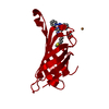


 PDBj
PDBj Assembly

 Mass: 590.240 Da / Num. of mol.: 1 / Source method: obtained synthetically / Formula: C27H38CuN6O3S
 Mass: 63.546 Da / Num. of mol.: 1 / Source method: obtained synthetically / Formula: Cu
 Mass: 92.094 Da / Num. of mol.: 1 / Source method: obtained synthetically / Formula: C3H8O3
 Mass: 18.015 Da / Num. of mol.: 93 / Source method: isolated from a natural source / Formula: H2O
 Sample preparation
 / Beamline: BL14-1 / Wavelength: 1.18 Å
 Processing