 Movie
Movie Controller


+ Open data

- Basic information
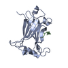
| Entry | Database: PDB / ID: 6anj | ||||||
|---|---|---|---|---|---|---|---|
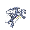
| Title | Synaptotagmin-7, C2A domain | ||||||
 Components Components | Synaptotagmin-7 | ||||||
 Keywords Keywords | PROTEIN BINDING / CALCIUM/PHOSPHOLIPID BINDING PROTEIN | ||||||
| Function / homology |  Function and homology information Function and homology informationcalcium ion regulated lysosome exocytosis / vesicle-mediated cholesterol transport / regulation of glucagon secretion / regulation of bone remodeling / phagosome-lysosome fusion / calcium-dependent activation of synaptic vesicle fusion / calcium ion-regulated exocytosis of neurotransmitter / regulation of calcium ion-dependent exocytosis / synaptic vesicle recycling / calcium ion sensor activity ...calcium ion regulated lysosome exocytosis / vesicle-mediated cholesterol transport / regulation of glucagon secretion / regulation of bone remodeling / phagosome-lysosome fusion / calcium-dependent activation of synaptic vesicle fusion / calcium ion-regulated exocytosis of neurotransmitter / regulation of calcium ion-dependent exocytosis / synaptic vesicle recycling / calcium ion sensor activity / short-term synaptic potentiation / positive regulation of calcium ion-dependent exocytosis / calcium-ion regulated exocytosis / vesicle fusion / calcium-dependent phospholipid binding / dense core granule / regulation of phagocytosis / early phagosome / plasma membrane repair / syntaxin binding / peroxisomal membrane / phosphatidylserine binding / clathrin binding / regulation of insulin secretion / regulation of dopamine secretion / regulation of synaptic vesicle endocytosis / detection of calcium ion / phagocytosis / vesicle-mediated transport / phosphatidylinositol-4,5-bisphosphate binding / axon terminus / hippocampal mossy fiber to CA3 synapse / SNARE binding / GABA-ergic synapse / phagocytic vesicle membrane / terminal bouton / synaptic vesicle / peroxisome / synaptic vesicle membrane / presynaptic membrane / calmodulin binding / lysosome / axon / lysosomal membrane / neuronal cell body / calcium ion binding / synapse / dendrite / glutamatergic synapse / plasma membrane / cytosol Similarity search - Function | ||||||
| Biological species |  | ||||||
| Method |  X-RAY DIFFRACTION / SYNCHROTRON / MOLECULAR REPLACEMENT / Resolution: 1.698 Å X-RAY DIFFRACTION / SYNCHROTRON / MOLECULAR REPLACEMENT / Resolution: 1.698 Å | ||||||
 Authors Authors | Tomchick, D.R. / Rizo, J. / Voleti, R. | ||||||
 Citation Citation | Journal: Proc. Natl. Acad. Sci. U.S.A. / Year: 2017 Title: Exceptionally tight membrane-binding may explain the key role of the synaptotagmin-7 C2A domain in asynchronous neurotransmitter release. Authors: Voleti, R. / Tomchick, D.R. / Sudhof, T.C. / Rizo, J. #1: Journal: PLoS ONE / Year: 2010Title: Structural and mutational analysis of functional differentiation between synaptotagmins-1 and -7. Authors: Xue, M. / Craig, T.K. / Shin, O.H. / Li, L. / Brautigam, C.A. / Tomchick, D.R. / Sudhof, T.C. / Rosenmund, C. / Rizo, J. | ||||||
| History |
|
- Structure visualization
| Structure viewer | Molecule: MolmilJmol/JSmol |
|---|
- Downloads & links
-Download
| PDBx/mmCIF format | 6anj.cif.gz | 105.5 KB | Display | PDBx/mmCIF format |
|---|---|---|---|---|
| PDB format | pdb6anj.ent.gz | 79.9 KB | Display | PDB format |
| PDBx/mmJSON format | 6anj.json.gz | Tree view | PDBx/mmJSON format | |
| Others |  Other downloads Other downloads |
-Validation report
| Arichive directory | https://data.pdbj.org/pub/pdb/validation_reports/an/6anjftp://data.pdbj.org/pub/pdb/validation_reports/an/6anj | HTTPS FTP |
|---|
-Related structure data
| Related structure data | 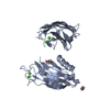 6ankC  2d8kS S: Starting model for refinement C: citing same article ( |
|---|---|
| Similar structure data |


-Links
 PDBj
PDBj


- Assembly
| Deposited unit | 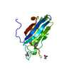
| ||||||||
|---|---|---|---|---|---|---|---|---|---|
| 1 |
| ||||||||
| Unit cell |
|
-Components
| #1: Protein | Mass: 16865.297 Da / Num. of mol.: 1 / Fragment: C2A domain (UNP residues 134-262) Source method: isolated from a genetically manipulated source Source: (gene. exp.)  | ||||||
|---|---|---|---|---|---|---|---|
| #2: Chemical |   Mass: 40.078 Da / Num. of mol.: 3 / Source method: obtained synthetically / Formula: Ca Mass: 40.078 Da / Num. of mol.: 3 / Source method: obtained synthetically / Formula: Ca#3: Chemical | 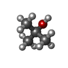  Mass: 74.122 Da / Num. of mol.: 3 / Source method: obtained synthetically / Formula: C4H10O Mass: 74.122 Da / Num. of mol.: 3 / Source method: obtained synthetically / Formula: C4H10O#4: Chemical | ChemComp-ACT / |   Mass: 59.044 Da / Num. of mol.: 1 / Source method: obtained synthetically / Formula: C2H3O2 Mass: 59.044 Da / Num. of mol.: 1 / Source method: obtained synthetically / Formula: C2H3O2#5: Water | ChemComp-HOH / |  Mass: 18.015 Da / Num. of mol.: 186 / Source method: isolated from a natural source / Formula: H2O Mass: 18.015 Da / Num. of mol.: 186 / Source method: isolated from a natural source / Formula: H2O |
-Experimental details
-Experiment
| Experiment | Method: X-RAY DIFFRACTION / Number of used crystals: 1 |
|---|
- Sample preparation
| Crystal | Density Matthews: 2.54 Å3/Da / Density % sol: 51.67 % |
|---|---|
| Crystal grow | Temperature: 293 K / Method: vapor diffusion, hanging drop / pH: 8.5 Details: 21% t-butanol, 0.1 M Tris, 0.1 M calcium chloride, 0.125 M KCl, 15% ethylene glycol |
-Data collection
| Diffraction | Mean temperature: 100 K |
|---|---|
| Diffraction source | Source: SYNCHROTRON / Site: APS  / Beamline: 19-ID / Wavelength: 0.97915 Å / Beamline: 19-ID / Wavelength: 0.97915 Å |
| Detector | Type: ADSC QUANTUM 315r / Detector: CCD / Date: Jun 14, 2014 / Details: monochromator |
| Radiation | Protocol: SINGLE WAVELENGTH / Monochromatic (M) / Laue (L): M / Scattering type: x-ray |
| Radiation wavelength | Wavelength: 0.97915 Å / Relative weight: 1 |
| Reflection | Resolution: 1.698→27.651 Å / Num. obs: 17195 / % possible obs: 99.7 % / Redundancy: 6.6 % / Biso Wilson estimate: 11.39 Å2 / Rmerge(I) obs: 0.054 / Rpim(I) all: 0.022 / Net I/σ(I): 34.6 |
| Reflection shell | Resolution: 1.7→1.73 Å / Redundancy: 4.1 % / Rmerge(I) obs: 0.985 / Mean I/σ(I) obs: 1.42 / CC1/2: 0.542 / Rpim(I) all: 0.528 / % possible all: 99.5 |
- Processing
| Software |
| |||||||||||||||||||||||||||||||||||||||||||||||||||||||||||||||||||||||||||||||||||||||||||||||||||||||||||||||||||||||||||||
|---|---|---|---|---|---|---|---|---|---|---|---|---|---|---|---|---|---|---|---|---|---|---|---|---|---|---|---|---|---|---|---|---|---|---|---|---|---|---|---|---|---|---|---|---|---|---|---|---|---|---|---|---|---|---|---|---|---|---|---|---|---|---|---|---|---|---|---|---|---|---|---|---|---|---|---|---|---|---|---|---|---|---|---|---|---|---|---|---|---|---|---|---|---|---|---|---|---|---|---|---|---|---|---|---|---|---|---|---|---|---|---|---|---|---|---|---|---|---|---|---|---|---|---|---|---|---|
| Refinement | Method to determine structure: MOLECULAR REPLACEMENT Starting model: 2D8K Resolution: 1.698→27.651 Å / SU ML: 0.16 / Cross valid method: FREE R-VALUE / σ(F): 1.35 / Phase error: 17.17
| |||||||||||||||||||||||||||||||||||||||||||||||||||||||||||||||||||||||||||||||||||||||||||||||||||||||||||||||||||||||||||||
| Solvent computation | Shrinkage radii: 0.9 Å / VDW probe radii: 1.11 Å | |||||||||||||||||||||||||||||||||||||||||||||||||||||||||||||||||||||||||||||||||||||||||||||||||||||||||||||||||||||||||||||
| Displacement parameters | Biso max: 84.68 Å2 / Biso mean: 17.4537 Å2 / Biso min: 4.32 Å2 | |||||||||||||||||||||||||||||||||||||||||||||||||||||||||||||||||||||||||||||||||||||||||||||||||||||||||||||||||||||||||||||
| Refinement step | Cycle: final / Resolution: 1.698→27.651 Å
| |||||||||||||||||||||||||||||||||||||||||||||||||||||||||||||||||||||||||||||||||||||||||||||||||||||||||||||||||||||||||||||
| Refine LS restraints |
| |||||||||||||||||||||||||||||||||||||||||||||||||||||||||||||||||||||||||||||||||||||||||||||||||||||||||||||||||||||||||||||
| LS refinement shell | Refine-ID: X-RAY DIFFRACTION / Rfactor Rfree error: 0 / Total num. of bins used: 12
| |||||||||||||||||||||||||||||||||||||||||||||||||||||||||||||||||||||||||||||||||||||||||||||||||||||||||||||||||||||||||||||
| Refinement TLS params. | Method: refined / Refine-ID: X-RAY DIFFRACTION
| |||||||||||||||||||||||||||||||||||||||||||||||||||||||||||||||||||||||||||||||||||||||||||||||||||||||||||||||||||||||||||||
| Refinement TLS group |
|
