regulation of white fat cell proliferation / tRNA demethylase activity / Reversal of alkylation damage by DNA dioxygenases / mRNA N6-methyladenine demethylase / mRNA N6-methyladenosine dioxygenase activity / regulation of respiratory system process / regulation of brown fat cell differentiation / regulation of lipid storage / broad specificity oxidative DNA demethylase activity / oxidative RNA demethylase activity ...regulation of white fat cell proliferation / tRNA demethylase activity / Reversal of alkylation damage by DNA dioxygenases / mRNA N6-methyladenine demethylase / mRNA N6-methyladenosine dioxygenase activity / regulation of respiratory system process / regulation of brown fat cell differentiation / regulation of lipid storage / broad specificity oxidative DNA demethylase activity / oxidative RNA demethylase activity / snRNA processing / RNA repair / DNA alkylation repair / Oxidoreductases; Acting on paired donors, with incorporation or reduction of molecular oxygen; With 2-oxoglutarate as one donor, and incorporation of one atom of oxygen into each donor / adipose tissue development / mRNA destabilization / regulation of multicellular organism growth / temperature homeostasis / ferrous iron binding / transferase activity / nuclear speck / nucleoplasm / nucleus / cytosol / cytoplasm Similarity search - Function
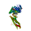
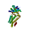
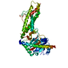
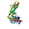
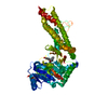
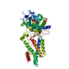
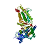
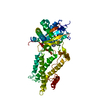
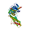
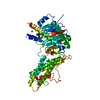
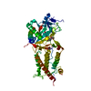
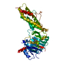
Alpha-ketoglutarate-dependentdioxygenaseFTO / Fat mass and obesity-associated protein
Mass: 56525.668 Da / Num. of mol.: 1 Source method: isolated from a genetically manipulated source Source: (gene. exp.) Homo sapiens (human) / Gene: FTO, KIAA1752 / Production host: Escherichia coli (E. coli) References: UniProt: Q9C0B1, Oxidoreductases; Acting on paired donors, with incorporation or reduction of molecular oxygen; With 2-oxoglutarate as one donor, and incorporation of one atom of oxygen into each donor
In the structure databanks used in Yorodumi, some data are registered as the other names, "COVID-19 virus" and "2019-nCoV". Here are the details of the virus and the list of structure data.
Jan 31, 2019. EMDB accession codes are about to change! (news from PDBe EMDB page)
EMDB accession codes are about to change! (news from PDBe EMDB page)
The allocation of 4 digits for EMDB accession codes will soon come to an end. Whilst these codes will remain in use, new EMDB accession codes will include an additional digit and will expand incrementally as the available range of codes is exhausted. The current 4-digit format prefixed with “EMD-” (i.e. EMD-XXXX) will advance to a 5-digit format (i.e. EMD-XXXXX), and so on. It is currently estimated that the 4-digit codes will be depleted around Spring 2019, at which point the 5-digit format will come into force.
The EM Navigator/Yorodumi systems omit the EMD- prefix.
Related info.:Q: What is EMD? / ID/Accession-code notation in Yorodumi/EM Navigator
Yorodumi is a browser for structure data from EMDB, PDB, SASBDB, etc.
This page is also the successor to EM Navigator detail page, and also detail information page/front-end page for Omokage search.
The word "yorodu" (or yorozu) is an old Japanese word meaning "ten thousand". "mi" (miru) is to see.
Related info.:EMDB / PDB / SASBDB / Comparison of 3 databanks / Yorodumi Search / Aug 31, 2016. New EM Navigator & Yorodumi / Yorodumi Papers / Jmol/JSmol / Function and homology information / Changes in new EM Navigator and Yorodumi
 Movie
Movie Controller


 Open data
 Basic information
 Components
 Keywords
 Function and homology information
 Homo sapiens (human)
 X-RAY DIFFRACTION /
  Authors
 Citation
 Structure visualization
 Downloads & links
 Other downloads
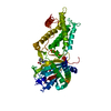


 PDBj
PDBj

 Assembly
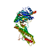


 Mass: 146.098 Da / Num. of mol.: 1 / Source method: obtained synthetically / Formula: C5H6O5
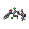
 Mass: 377.221 Da / Num. of mol.: 1 / Source method: obtained synthetically / Formula: C18H14Cl2N2O3 / Feature type: SUBJECT OF INVESTIGATION
 Mass: 18.015 Da / Num. of mol.: 153 / Source method: isolated from a natural source / Formula: H2O
 Sample preparation
 / Beamline: BL18U1 / Wavelength: 0.9735 Å
 Processing