regulation of white fat cell proliferation / tRNA demethylase activity / Reversal of alkylation damage by DNA dioxygenases / mRNA N6-methyladenine demethylase / mRNA N6-methyladenosine dioxygenase activity / regulation of respiratory system process / regulation of lipid storage / regulation of brown fat cell differentiation / broad specificity oxidative DNA demethylase activity / oxidative RNA demethylase activity ...regulation of white fat cell proliferation / tRNA demethylase activity / Reversal of alkylation damage by DNA dioxygenases / mRNA N6-methyladenine demethylase / mRNA N6-methyladenosine dioxygenase activity / regulation of respiratory system process / regulation of lipid storage / regulation of brown fat cell differentiation / broad specificity oxidative DNA demethylase activity / oxidative RNA demethylase activity / snRNA processing / RNA repair / DNA alkylation repair / Oxidoreductases; Acting on paired donors, with incorporation or reduction of molecular oxygen; With 2-oxoglutarate as one donor, and incorporation of one atom of oxygen into each donor / mRNA destabilization / temperature homeostasis / regulation of multicellular organism growth / adipose tissue development / ferrous iron binding / transferase activity / nuclear speck / nucleoplasm / nucleus / cytoplasm / cytosol Similarity search - Function
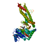
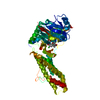
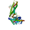
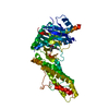
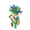
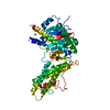
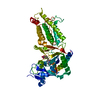
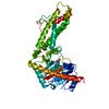
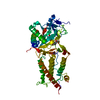
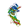
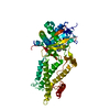
Alpha-ketoglutarate-dependentdioxygenaseFTO / Fat mass and obesity-associated protein
Mass: 54933.965 Da / Num. of mol.: 1 / Fragment: UNP residues 32-505 Source method: isolated from a genetically manipulated source Source: (gene. exp.) Homo sapiens (human) / Gene: FTO, KIAA1752 / Production host: Escherichia coli (E. coli) References: UniProt: Q9C0B1, Oxidoreductases; Acting on paired donors, with incorporation or reduction of molecular oxygen; With 2-oxoglutarate as one donor, and incorporation of one atom of oxygen into each donor
Type: APEX II CCD / Detector: CCD / Date: Jan 12, 2015
Radiation
Protocol: SINGLE WAVELENGTH / Monochromatic (M) / Laue (L): M / Scattering type: x-ray
Radiation wavelength
Wavelength: 0.9735 Å / Relative weight: 1
Reflection
Resolution: 2.45→30 Å / Num. obs: 23094 / % possible obs: 100 % / Redundancy: 10.4 % / Net I/σ(I): 27.6
-
Processing
Software
Name
Version
Classification
REFMAC
5.7.0029
refinement
HKL-3000
datareduction
HKL-3000
datascaling
PHASER
phasing
Refinement
Method to determine structure: MOLECULAR REPLACEMENT / Resolution: 2.45→28.91 Å / Cor.coef. Fo:Fc: 0.956 / Cor.coef. Fo:Fc free: 0.927 / SU B: 8.715 / SU ML: 0.197 / Cross valid method: THROUGHOUT / ESU R: 0.358 / ESU R Free: 0.266 / Stereochemistry target values: MAXIMUM LIKELIHOOD Details: HYDROGENS HAVE BEEN ADDED IN THE RIDING POSITIONS. SF FILE CONTAINS FRIEDEL PAIRS UNDER I/F_MINUS AND I/F_PLUS COLUMNS.
Rfactor
Num. reflection
% reflection
Selection details
Rfree
0.26257
1181
5.1 %
RANDOM
Rwork
0.20608
-
-
-
obs
0.20889
21848
99.58 %
-
Solvent computation
Ion probe radii: 0.8 Å / Shrinkage radii: 0.8 Å / VDW probe radii: 1.2 Å / Solvent model: MASK
 Movie
Movie Controller

 Yorodumi
 Open data
 Basic information
 Components
 Keywords
 Function and homology information
 Homo sapiens (human)
 X-RAY DIFFRACTION /
  Authors
 Citation
 Structure visualization
 Downloads & links
 Other downloads

 PDBj
PDBj

 Assembly
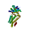


 Mass: 146.098 Da / Num. of mol.: 1 / Source method: obtained synthetically / Formula: C5H6O5
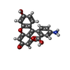
 Mass: 347.321 Da / Num. of mol.: 1 / Source method: obtained synthetically / Formula: C20H13NO5
 Mass: 54.938 Da / Num. of mol.: 1 / Source method: obtained synthetically / Formula: Mn
 Mass: 18.015 Da / Num. of mol.: 52 / Source method: isolated from a natural source / Formula: H2O
 Sample preparation
 / Beamline: BL19U1 / Wavelength: 0.9735 Å
 Processing