 Movie
Movie Controller

[English] 日本語
 Yorodumi
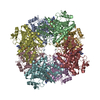
- PDB-6acf: structure of leucine dehydrogenase from Geobacillus stearothermop... -
+ Open data

- Basic information
| Entry | Database: PDB / ID: 6acf | ||||||
|---|---|---|---|---|---|---|---|
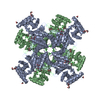
| Title | structure of leucine dehydrogenase from Geobacillus stearothermophilus by cryo-EM | ||||||
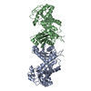
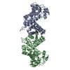
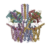
 Components Components | Leucine dehydrogenase | ||||||
 Keywords Keywords | OXIDOREDUCTASE / LEUCINE DEHYDROGENSE / NAD/LEUCINE BINDING / APO FORM | ||||||
| Function / homology |  Function and homology information Function and homology informationoxidoreductase activity, acting on the CH-NH2 group of donors, NAD or NADP as acceptor / amino acid metabolic process / nucleotide binding Similarity search - Function | ||||||
| Biological species |   Geobacillus stearothermophilus 10 (bacteria) Geobacillus stearothermophilus 10 (bacteria) | ||||||
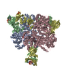
| Method | ELECTRON MICROSCOPY / single particle reconstruction / cryo EM / Resolution: 3 Å | ||||||
 Authors Authors | Yamaguchi, H. / Kamegawa, A. / Nakata, K. / Kashiwagi, T. / Mizukoshi, T. / Fujiyoshi, Y. / Tani, K. | ||||||
| Funding support |  Japan, 1items Japan, 1items
| ||||||
 Citation Citation | Journal: J Struct Biol / Year: 2019 Title: Structural insights into thermostabilization of leucine dehydrogenase from its atomic structure by cryo-electron microscopy. Authors: Hiroki Yamaguchi / Akiko Kamegawa / Kunio Nakata / Tatsuki Kashiwagi / Toshimi Mizukoshi / Yoshinori Fujiyoshi / Kazutoshi Tani / Abstract: Leucine dehydrogenase (LDH, EC 1.4.1.9) is a NAD-dependent oxidoreductase that catalyzes the deamination of branched-chain l-amino acids (BCAAs). LDH of Geobacillus stearothermophilus (GstLDH) is a ...Leucine dehydrogenase (LDH, EC 1.4.1.9) is a NAD-dependent oxidoreductase that catalyzes the deamination of branched-chain l-amino acids (BCAAs). LDH of Geobacillus stearothermophilus (GstLDH) is a highly thermostable enzyme that has been applied for the quantification or production of BCAAs. Here the cryo-electron microscopy (cryo-EM) structures of apo and NAD-bound LDH are reported at 3.0 and 3.2 Å resolution, respectively. On comparing the structures, the two overall structures are almost identical, but it was observed that the partial conformational change was triggered by the interaction between Ser147 and the nicotinamide moiety of NAD. NAD binding also enhanced the strength of oligomerization interfaces formed by the core domains. Such additional interdomain interaction is in good agreement with our experimental results showing that the residual activity of NAD-bound form was approximately three times higher than that of the apo form after incubation at 80 °C. In addition, sequence comparison of three structurally known LDHs indicated a set of candidates for site-directed mutagenesis to improve thermostability. Subsequent mutation analysis actually revealed that non-conserved residues, including Ala94, Tyr127, and the C-terminal region, are crucial for oligomeric thermostability. | ||||||
| History |
|
- Structure visualization
| Movie |
Movie viewer |
|---|---|
| Structure viewer | Molecule: MolmilJmol/JSmol |
- Downloads & links
-Download
| PDBx/mmCIF format | 6acf.cif.gz | 514.2 KB | Display | PDBx/mmCIF format |
|---|---|---|---|---|
| PDB format | pdb6acf.ent.gz | 430.3 KB | Display | PDB format |
| PDBx/mmJSON format | 6acf.json.gz | Tree view | PDBx/mmJSON format | |
| Others |  Other downloads Other downloads |
-Validation report
| Arichive directory | https://data.pdbj.org/pub/pdb/validation_reports/ac/6acfftp://data.pdbj.org/pub/pdb/validation_reports/ac/6acf | HTTPS FTP |
|---|
-Related structure data
| Related structure data | 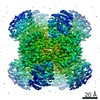 9590MC 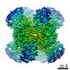 9592C 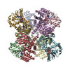 6achC M: map data used to model this data C: citing same article ( |
|---|---|
| Similar structure data |
-Links
 PDBj
PDBj- Assembly
| Deposited unit | 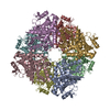
|
|---|---|
| 1 |
|
-Components
| #1: Protein | Mass: 40584.020 Da / Num. of mol.: 8 Source method: isolated from a genetically manipulated source Source: (gene. exp.) Geobacillus stearothermophilus 10 (bacteria)Gene: GT50_15010 / Production host: |
|---|
-Experimental details
-Experiment
| Experiment | Method: ELECTRON MICROSCOPY |
|---|---|
| EM experiment | Aggregation state: PARTICLE / 3D reconstruction method: single particle reconstruction |
- Sample preparation
| Component | Name: The apo form of Leucine dehydrogenase / Type: COMPLEX / Entity ID: all / Source: RECOMBINANT |
|---|---|
| Molecular weight | Value: 0.305 MDa / Experimental value: YES |
| Source (natural) | Organism: Geobacillus stearothermophilus 10 (bacteria) |
| Source (recombinant) | Organism: |
| Buffer solution | pH: 10.5 |
| Specimen | Conc.: 0.5 mg/ml / Embedding applied: NO / Shadowing applied: NO / Staining applied: NO / Vitrification applied: YES |
| Specimen support | Grid material: MOLYBDENUM / Grid mesh size: 200 divisions/in. / Grid type: Quantifoil R2/2 |
| Vitrification | Cryogen name: ETHANE |
- Electron microscopy imaging
| Microscopy | Model: JEOL KYOTO-3000SFF |
|---|---|
| Electron gun | Electron source:  FIELD EMISSION GUN / Accelerating voltage: 300 kV / Illumination mode: FLOOD BEAM FIELD EMISSION GUN / Accelerating voltage: 300 kV / Illumination mode: FLOOD BEAM |
| Electron lens | Mode: BRIGHT FIELD / Cs: 1.6 mm |
| Image recording | Electron dose: 2.7 e/Å2 / Detector mode: SUPER-RESOLUTION / Film or detector model: GATAN K2 SUMMIT (4k x 4k) |
- Processing
| Software | Name: PHENIX / Version: 1.12_2829: / Classification: refinement | ||||||||||||||||||||||||||||||||
|---|---|---|---|---|---|---|---|---|---|---|---|---|---|---|---|---|---|---|---|---|---|---|---|---|---|---|---|---|---|---|---|---|---|
| EM software |
| ||||||||||||||||||||||||||||||||
| CTF correction | Type: PHASE FLIPPING ONLY | ||||||||||||||||||||||||||||||||
| Symmetry | Point symmetry: D4 (2x4 fold dihedral) | ||||||||||||||||||||||||||||||||
| 3D reconstruction | Resolution: 3 Å / Resolution method: FSC 0.143 CUT-OFF / Num. of particles: 132800 / Symmetry type: POINT | ||||||||||||||||||||||||||||||||
| Refine LS restraints |
|
