Movie
Movie Controller

[English] 日本語
 Yorodumi
- EMDB-9592: Structure of NAD+-bound leucine dehydrogenase from Geobacillus st... -
+ Open data

- Basic information
| Entry | Database: EMDB / ID: EMD-9592 | |||||||||
|---|---|---|---|---|---|---|---|---|---|---|
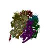
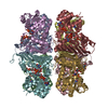
| Title | Structure of NAD+-bound leucine dehydrogenase from Geobacillus stearothermophilus by cryo-EM | |||||||||
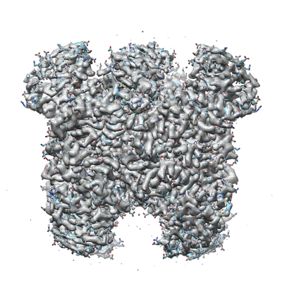
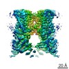
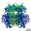
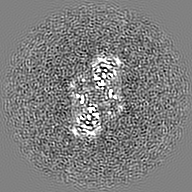
 Map data Map data | The holo form of GstLDH | |||||||||
 Sample Sample |
| |||||||||
 Keywords Keywords | LEUCINE DEHYDROGENSE / NAD/LEUCINE BINDING / HOLO FORM / OXIDOREDUCTASE | |||||||||
| Function / homology |  Function and homology information Function and homology informationoxidoreductase activity, acting on the CH-NH2 group of donors, NAD or NADP as acceptor / amino acid metabolic process / nucleotide binding Similarity search - Function | |||||||||
| Biological species |   Geobacillus stearothermophilus 10 (bacteria) Geobacillus stearothermophilus 10 (bacteria) | |||||||||
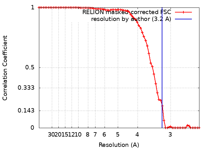
| Method | single particle reconstruction / cryo EM / Resolution: 3.2 Å | |||||||||
 Authors Authors | Yamaguchi H / Kamegawa A / Nakata K / Kashiwagi T / Mizukoshi T / Fujiyoshi Y / Tani K | |||||||||
 Citation Citation | Journal: J Struct Biol / Year: 2019 Title: Structural insights into thermostabilization of leucine dehydrogenase from its atomic structure by cryo-electron microscopy. Authors: Hiroki Yamaguchi / Akiko Kamegawa / Kunio Nakata / Tatsuki Kashiwagi / Toshimi Mizukoshi / Yoshinori Fujiyoshi / Kazutoshi Tani /  Abstract: Leucine dehydrogenase (LDH, EC 1.4.1.9) is a NAD-dependent oxidoreductase that catalyzes the deamination of branched-chain l-amino acids (BCAAs). LDH of Geobacillus stearothermophilus (GstLDH) is a ...Leucine dehydrogenase (LDH, EC 1.4.1.9) is a NAD-dependent oxidoreductase that catalyzes the deamination of branched-chain l-amino acids (BCAAs). LDH of Geobacillus stearothermophilus (GstLDH) is a highly thermostable enzyme that has been applied for the quantification or production of BCAAs. Here the cryo-electron microscopy (cryo-EM) structures of apo and NAD-bound LDH are reported at 3.0 and 3.2 Å resolution, respectively. On comparing the structures, the two overall structures are almost identical, but it was observed that the partial conformational change was triggered by the interaction between Ser147 and the nicotinamide moiety of NAD. NAD binding also enhanced the strength of oligomerization interfaces formed by the core domains. Such additional interdomain interaction is in good agreement with our experimental results showing that the residual activity of NAD-bound form was approximately three times higher than that of the apo form after incubation at 80 °C. In addition, sequence comparison of three structurally known LDHs indicated a set of candidates for site-directed mutagenesis to improve thermostability. Subsequent mutation analysis actually revealed that non-conserved residues, including Ala94, Tyr127, and the C-terminal region, are crucial for oligomeric thermostability. | |||||||||
| History |
|
- Structure visualization
| Movie |
Movie viewer |
|---|---|
| Structure viewer | EM map: SurfViewMolmilJmol/JSmol |
| Supplemental images |
- Downloads & links
-EMDB archive
| Map data | emd_9592.map.gz | 25.1 MB | EMDB map data format | |
|---|---|---|---|---|
| Header (meta data) | emd-9592-v30.xmlemd-9592.xml | 11.2 KB 11.2 KB | Display Display | EMDB header |
| FSC (resolution estimation) | emd_9592_fsc.xml | 6.7 KB | Display | FSC data file |
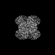
| Images |  emd_9592.png emd_9592.png | 208.2 KB | ||
| Filedesc metadata | emd-9592.cif.gz | 5.6 KB | ||
| Archive directory |  http://ftp.pdbj.org/pub/emdb/structures/EMD-9592ftp://ftp.pdbj.org/pub/emdb/structures/EMD-9592 http://ftp.pdbj.org/pub/emdb/structures/EMD-9592ftp://ftp.pdbj.org/pub/emdb/structures/EMD-9592 | HTTPS FTP |
-Related structure data
| Related structure data |  6achMC  9590C 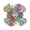 6acfC M: atomic model generated by this map C: citing same article ( |
|---|---|
| Similar structure data |

-Links
| EMDB pages | EMDB (EBI/PDBe) / EMDataResource |
|---|
-Map
| File | Download / File: emd_9592.map.gz / Format: CCP4 / Size: 27 MB / Type: IMAGE STORED AS FLOATING POINT NUMBER (4 BYTES) | ||||||||||||||||||||||||||||||||||||||||||||||||||||||||||||
|---|---|---|---|---|---|---|---|---|---|---|---|---|---|---|---|---|---|---|---|---|---|---|---|---|---|---|---|---|---|---|---|---|---|---|---|---|---|---|---|---|---|---|---|---|---|---|---|---|---|---|---|---|---|---|---|---|---|---|---|---|---|
| Annotation | The holo form of GstLDH | ||||||||||||||||||||||||||||||||||||||||||||||||||||||||||||
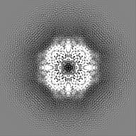
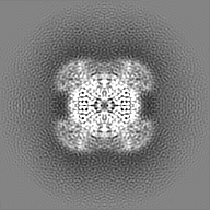
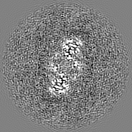
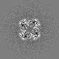
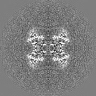
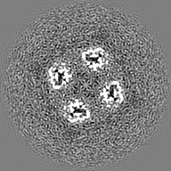
| Projections & slices | Image control
Images are generated by Spider. | ||||||||||||||||||||||||||||||||||||||||||||||||||||||||||||
| Voxel size | X=Y=Z: 1.232 Å | ||||||||||||||||||||||||||||||||||||||||||||||||||||||||||||
| Density |
| ||||||||||||||||||||||||||||||||||||||||||||||||||||||||||||
| Symmetry | Space group: 1 | ||||||||||||||||||||||||||||||||||||||||||||||||||||||||||||
| Details | EMDB XML:
CCP4 map header:
| ||||||||||||||||||||||||||||||||||||||||||||||||||||||||||||
 Z (Sec.)
 Y (Row.)
 X (Col.)


-Supplemental data
- Sample components
-Entire : Binary complex of leucine dehydrogenase with NAD+
| Entire | Name: Binary complex of leucine dehydrogenase with NAD+ |
|---|---|
| Components |
|
-Supramolecule #1: Binary complex of leucine dehydrogenase with NAD+
| Supramolecule | Name: Binary complex of leucine dehydrogenase with NAD+ / type: complex / ID: 1 / Parent: 0 / Macromolecule list: #1 |
|---|---|
| Source (natural) | Organism: Geobacillus stearothermophilus 10 (bacteria) |
| Molecular weight | Theoretical: 0.305 kDa/nm |
-Macromolecule #1: Leucine dehydrogenase
| Macromolecule | Name: Leucine dehydrogenase / type: protein_or_peptide / ID: 1 / Number of copies: 8 / Enantiomer: LEVO |
|---|---|
| Source (natural) | Organism: Geobacillus stearothermophilus 10 (bacteria) |
| Molecular weight | Theoretical: 40.58402 KDa |
| Recombinant expression | Organism: |
| Sequence | String: MELFQYMEKY DYEQVLFCQD KESGLKAIIV IHDTTLGPAL GGTRMWMYNS EEEALEDALR LARGMTYKNA AAGLNLGGGK TVIIGDPRK DKNEAMFRAF GRFIQGLNGR YITAEDVGTT VADMDIIYQE TDYVTGISPE FGSSGNPSPA TAYGVYRGMK A AAKEAFGS ...String: MELFQYMEKY DYEQVLFCQD KESGLKAIIV IHDTTLGPAL GGTRMWMYNS EEEALEDALR LARGMTYKNA AAGLNLGGGK TVIIGDPRK DKNEAMFRAF GRFIQGLNGR YITAEDVGTT VADMDIIYQE TDYVTGISPE FGSSGNPSPA TAYGVYRGMK A AAKEAFGS DSLEGKVVAV QGVGNVAYHL CRHLHEEGAK LIVTDINKEA VARAVEEFGA KAVDPNDIYG VECDIFAPCA LG GIINDQT IPQLKAKVIA GSANNQLKEP RHGDMIHEMG IVYAPDYVIN AGGVINVADE LYGYNRERAM KKIEQIYDNI EKV FAIAKR DNIPTYVAAD RMAEERIETM RKARSQFLQN GHHILSRRRA R UniProtKB: Leucine dehydrogenase |
-Macromolecule #2: NICOTINAMIDE-ADENINE-DINUCLEOTIDE
| Macromolecule | Name: NICOTINAMIDE-ADENINE-DINUCLEOTIDE / type: ligand / ID: 2 / Number of copies: 8 / Formula: NAD |
|---|---|
| Molecular weight | Theoretical: 663.425 Da |
| Chemical component information |  ChemComp-NAD: |
-Experimental details
-Structure determination
| Method | cryo EM |
|---|---|
 Processing Processing | single particle reconstruction |
| Aggregation state | particle |
-Sample preparation
| Concentration | 0.5 mg/mL |
|---|---|
| Buffer | pH: 10.5 |
| Grid | Model: Quantifoil R2/2 / Material: MOLYBDENUM / Mesh: 200 |
| Vitrification | Cryogen name: ETHANE |
- Electron microscopy
| Microscope | JEOL KYOTO-3000SFF |
|---|---|
| Image recording | Film or detector model: GATAN K2 SUMMIT (4k x 4k) / Detector mode: SUPER-RESOLUTION / Average electron dose: 2.7 e/Å2 |
| Electron beam | Acceleration voltage: 300 kV / Electron source:  FIELD EMISSION GUN FIELD EMISSION GUN |
| Electron optics | Illumination mode: FLOOD BEAM / Imaging mode: BRIGHT FIELD |