 Movie
Movie Controller


+ Open data

- Basic information
| Entry | Database: PDB / ID: 7byn | |||||||||||||||||||||||||||||||||||||||||||||
|---|---|---|---|---|---|---|---|---|---|---|---|---|---|---|---|---|---|---|---|---|---|---|---|---|---|---|---|---|---|---|---|---|---|---|---|---|---|---|---|---|---|---|---|---|---|---|
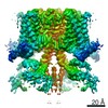
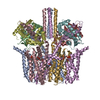
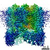
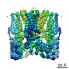
| Title | Cryo-EM structure of human KCNQ4 with linopirdine | |||||||||||||||||||||||||||||||||||||||||||||
 Components Components |
| |||||||||||||||||||||||||||||||||||||||||||||
 Keywords Keywords | MEMBRANE PROTEIN / KCNQ / Channel / Calmodulin / PIP2 / linopirdine | |||||||||||||||||||||||||||||||||||||||||||||
| Function / homology |  Function and homology information Function and homology informationtransporter inhibitor activity / : / type 3 metabotropic glutamate receptor binding / Voltage gated Potassium channels / Sensory processing of sound by outer hair cells of the cochlea / Sensory processing of sound by inner hair cells of the cochlea / inner ear morphogenesis / negative regulation of high voltage-gated calcium channel activity / response to corticosterone / regulation of synaptic vesicle exocytosis ...transporter inhibitor activity / : / type 3 metabotropic glutamate receptor binding / Voltage gated Potassium channels / Sensory processing of sound by outer hair cells of the cochlea / Sensory processing of sound by inner hair cells of the cochlea / inner ear morphogenesis / negative regulation of high voltage-gated calcium channel activity / response to corticosterone / regulation of synaptic vesicle exocytosis / negative regulation of calcium ion export across plasma membrane / regulation of cardiac muscle cell action potential / presynaptic endocytosis / calcineurin-mediated signaling / nitric-oxide synthase binding / regulation of cell communication by electrical coupling involved in cardiac conduction / adenylate cyclase binding / protein phosphatase activator activity / regulation of synaptic vesicle endocytosis / voltage-gated potassium channel activity / potassium channel activity / detection of calcium ion / postsynaptic cytosol / regulation of cardiac muscle contraction / catalytic complex / phosphatidylinositol 3-kinase binding / presynaptic cytosol / regulation of release of sequestered calcium ion into cytosol by sarcoplasmic reticulum / titin binding / regulation of cardiac muscle contraction by regulation of the release of sequestered calcium ion / regulation of calcium-mediated signaling / voltage-gated potassium channel complex / calcium channel complex / potassium ion transmembrane transport / substantia nigra development / regulation of heart rate / basal plasma membrane / calyx of Held / nitric-oxide synthase regulator activity / adenylate cyclase activator activity / bioluminescence / protein serine/threonine kinase activator activity / regulation of cytokinesis / spindle microtubule / response to amphetamine / sarcomere / calcium channel regulator activity / generation of precursor metabolites and energy / potassium ion transport / sensory perception of sound / response to calcium ion / G2/M transition of mitotic cell cycle / Schaffer collateral - CA1 synapse / mitochondrial membrane / spindle pole / calcium-dependent protein binding / long-term synaptic potentiation / myelin sheath / synaptic vesicle membrane / growth cone / sperm midpiece / vesicle / transmembrane transporter binding / G protein-coupled receptor signaling pathway / protein domain specific binding / calcium ion binding / centrosome / protein kinase binding / protein-containing complex / nucleus / plasma membrane / cytoplasm Similarity search - Function | |||||||||||||||||||||||||||||||||||||||||||||
| Biological species |   Aequorea victoria (jellyfish) Aequorea victoria (jellyfish) Homo sapiens (human) Homo sapiens (human) | |||||||||||||||||||||||||||||||||||||||||||||
| Method | ELECTRON MICROSCOPY / single particle reconstruction / cryo EM / Resolution: 3.3 Å | |||||||||||||||||||||||||||||||||||||||||||||
 Authors Authors | Shen, H. / Li, T. / Yue, Z. | |||||||||||||||||||||||||||||||||||||||||||||
 Citation Citation | Journal: Mol Cell / Year: 2021 Title: Structural Basis for the Modulation of Human KCNQ4 by Small-Molecule Drugs. Authors: Tian Li / Kun Wu / Zhenlei Yue / Yifei Wang / Fan Zhang / Huaizong Shen /  Abstract: Among the five KCNQ channels, also known as the K7 voltage-gated potassium (K) channels, KCNQ2-KCNQ5 control neuronal excitability. Dysfunctions of KCNQ2-KCNQ5 are associated with neurological ...Among the five KCNQ channels, also known as the K7 voltage-gated potassium (K) channels, KCNQ2-KCNQ5 control neuronal excitability. Dysfunctions of KCNQ2-KCNQ5 are associated with neurological disorders such as epilepsy, deafness, and neuropathic pain. Here, we report the cryoelectron microscopy (cryo-EM) structures of human KCNQ4 and its complexes with the opener retigabine or the blocker linopirdine at overall resolutions of 2.5, 3.1, and 3.3 Å, respectively. In all structures, a phosphatidylinositol 4,5-bisphosphate (PIP) molecule inserts its head group into a cavity within each voltage-sensing domain (VSD), revealing an unobserved binding mode for PIP. Retigabine nestles in each fenestration, inducing local shifts. Instead of staying within the central pore, linopirdine resides in a cytosolic cavity underneath the inner gate. Electrophysiological analyses of various mutants corroborated the structural observations. Our studies reveal the molecular basis for the modulatory mechanism of neuronal KCNQ channels and provide a framework for structure-facilitated drug discovery targeting these important channels. | |||||||||||||||||||||||||||||||||||||||||||||
| History |
|
- Structure visualization
| Movie |
Movie viewer |
|---|---|
| Structure viewer | Molecule: MolmilJmol/JSmol |
- Downloads & links
-Download
| PDBx/mmCIF format | 7byn.cif.gz | 429.7 KB | Display | PDBx/mmCIF format |
|---|---|---|---|---|
| PDB format | pdb7byn.ent.gz | 321.8 KB | Display | PDB format |
| PDBx/mmJSON format | 7byn.json.gz | Tree view | PDBx/mmJSON format | |
| Others |  Other downloads Other downloads |
-Validation report
| Arichive directory | https://data.pdbj.org/pub/pdb/validation_reports/by/7bynftp://data.pdbj.org/pub/pdb/validation_reports/by/7byn | HTTPS FTP |
|---|
-Related structure data
| Related structure data | 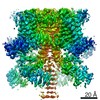 30246MC  7bylC  7bymC M: map data used to model this data C: citing same article ( |
|---|---|
| Similar structure data |


-Links
 PDBj
PDBj


- Assembly
| Deposited unit | 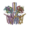
|
|---|---|
| 1 |
|
-Components
| #1: Protein | Mass: 109327.836 Da / Num. of mol.: 4 / Mutation: F64L/S65T/K107T/A206K/H231L Source method: isolated from a genetically manipulated source Source: (gene. exp.) Aequorea victoria (jellyfish), (gene. exp.) Homo sapiens (human)Gene: GFP, KCNQ4 / Production host: Homo sapiens (human) / References: UniProt: P42212, UniProt: P56696#2: Protein | Mass: 16852.545 Da / Num. of mol.: 4 Source method: isolated from a genetically manipulated source Source: (gene. exp.) Homo sapiens (human) / Gene: CALM3, CALML2, CAM3, CAMC, CAMIII / Production host: Homo sapiens (human) / References: UniProt: P0DP25#3: Chemical | ChemComp-PT5 / [(   Mass: 1047.088 Da / Num. of mol.: 4 / Source method: obtained synthetically / Formula: C47H85O19P3 / Feature type: SUBJECT OF INVESTIGATION / Comment: phospholipid*YM Mass: 1047.088 Da / Num. of mol.: 4 / Source method: obtained synthetically / Formula: C47H85O19P3 / Feature type: SUBJECT OF INVESTIGATION / Comment: phospholipid*YM#4: Chemical |   Mass: 39.098 Da / Num. of mol.: 3 / Source method: obtained synthetically / Formula: K Mass: 39.098 Da / Num. of mol.: 3 / Source method: obtained synthetically / Formula: K#5: Chemical | ChemComp-FCC / |   Mass: 391.464 Da / Num. of mol.: 1 / Source method: obtained synthetically / Formula: C26H21N3O / Feature type: SUBJECT OF INVESTIGATION Mass: 391.464 Da / Num. of mol.: 1 / Source method: obtained synthetically / Formula: C26H21N3O / Feature type: SUBJECT OF INVESTIGATIONHas ligand of interest | Y | Has protein modification | N | |
|---|
-Experimental details
-Experiment
| Experiment | Method: ELECTRON MICROSCOPY |
|---|---|
| EM experiment | Aggregation state: PARTICLE / 3D reconstruction method: single particle reconstruction |
- Sample preparation
| Component | Name: Complex of human KCNQ4 and linopirdine / Type: COMPLEX / Entity ID: #1-#2 / Source: RECOMBINANT |
|---|---|
| Source (natural) | Organism: Homo sapiens (human) |
| Source (recombinant) | Organism: Homo sapiens (human) |
| Buffer solution | pH: 8 |
| Specimen | Embedding applied: NO / Shadowing applied: NO / Staining applied: NO / Vitrification applied: YES |
| Vitrification | Cryogen name: ETHANE |
- Electron microscopy imaging
| Experimental equipment |  Model: Titan Krios / Image courtesy: FEI Company |
|---|---|
| Microscopy | Model: FEI TITAN KRIOS |
| Electron gun | Electron source:  FIELD EMISSION GUN / Accelerating voltage: 300 kV / Illumination mode: FLOOD BEAM FIELD EMISSION GUN / Accelerating voltage: 300 kV / Illumination mode: FLOOD BEAM |
| Electron lens | Mode: BRIGHT FIELD |
| Image recording | Electron dose: 50 e/Å2 / Film or detector model: GATAN K3 (6k x 4k) |
- Processing
| Software | Name: PHENIX / Version: 1.17.1_3660: / Classification: refinement | ||||||||||||||||||||||||
|---|---|---|---|---|---|---|---|---|---|---|---|---|---|---|---|---|---|---|---|---|---|---|---|---|---|
| EM software | Name: PHENIX / Category: model refinement | ||||||||||||||||||||||||
| CTF correction | Type: NONE | ||||||||||||||||||||||||
| 3D reconstruction | Resolution: 3.3 Å / Resolution method: FSC 0.143 CUT-OFF / Num. of particles: 229040 / Symmetry type: POINT | ||||||||||||||||||||||||
| Refine LS restraints |
|
