[English] 日本語
 Yorodumi
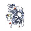
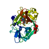
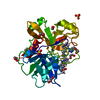
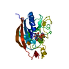
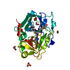
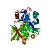
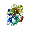
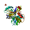
- PDB-6a8g: The crystal structure of muPAin-1-IG in complex with muPA-SPD at pH8.5 -
+ Open data
- Basic information
| Entry | Database: PDB / ID: 6a8g | ||||||||||||
|---|---|---|---|---|---|---|---|---|---|---|---|---|---|
| Title | The crystal structure of muPAin-1-IG in complex with muPA-SPD at pH8.5 | ||||||||||||
 Components Components |
| ||||||||||||
 Keywords Keywords | HYDROLASE INHIBITOR/HYDROLASE / Peptides inhibitor / muPA / serine protease / HYDROLASE / HYDROLASE INHIBITOR-HYDROLASE complex | ||||||||||||
| Function / homology |  Function and homology information Function and homology informationDissolution of Fibrin Clot / u-plasminogen activator / regulation of smooth muscle cell-matrix adhesion / urokinase plasminogen activator signaling pathway / regulation of plasminogen activation / regulation of fibrinolysis / protein complex involved in cell-matrix adhesion / negative regulation of plasminogen activation / regulation of smooth muscle cell migration / serine-type endopeptidase complex ...Dissolution of Fibrin Clot / u-plasminogen activator / regulation of smooth muscle cell-matrix adhesion / urokinase plasminogen activator signaling pathway / regulation of plasminogen activation / regulation of fibrinolysis / protein complex involved in cell-matrix adhesion / negative regulation of plasminogen activation / regulation of smooth muscle cell migration / serine-type endopeptidase complex / smooth muscle cell migration / plasminogen activation / regulation of cell adhesion mediated by integrin / negative regulation of fibrinolysis / regulation of cell adhesion / serine protease inhibitor complex / fibrinolysis / Neutrophil degranulation / regulation of cell population proliferation / response to hypoxia / positive regulation of cell migration / external side of plasma membrane / serine-type endopeptidase activity / extracellular space Similarity search - Function | ||||||||||||
| Biological species |  Phage display vector pTDisp (others) | ||||||||||||
| Method |  X-RAY DIFFRACTION / X-RAY DIFFRACTION /  SYNCHROTRON / SYNCHROTRON /  MOLECULAR REPLACEMENT / MOLECULAR REPLACEMENT /  molecular replacement / Resolution: 2.53 Å molecular replacement / Resolution: 2.53 Å | ||||||||||||
 Authors Authors | Wang, D. / Yang, Y.S. / Jiang, L.G. / Huang, M.D. / Li, J.Y. / Andreasen, P.A. / Xu, P. / Chen, Z. | ||||||||||||
| Funding support |  China, 3items China, 3items
| ||||||||||||
 Citation Citation |  Journal: J.Med.Chem. / Year: 2019 Journal: J.Med.Chem. / Year: 2019Title: Suppression of Tumor Growth and Metastases by Targeted Intervention in Urokinase Activity with Cyclic Peptides. Authors: Wang, D. / Yang, Y. / Jiang, L. / Wang, Y. / Li, J. / Andreasen, P.A. / Chen, Z. / Huang, M. / Xu, P. | ||||||||||||
| History |
|
- Structure visualization
| Structure viewer | Molecule:  Molmil Molmil Jmol/JSmol Jmol/JSmol |
|---|
- Downloads & links
- Download
| PDBx/mmCIF format |  6a8g.cif.gz 6a8g.cif.gz | 214.9 KB | Display |  PDBx/mmCIF format PDBx/mmCIF format |
|---|---|---|---|---|
| PDB format |  pdb6a8g.ent.gz pdb6a8g.ent.gz | 173.9 KB | Display |  PDB format PDB format |
| PDBx/mmJSON format |  6a8g.json.gz 6a8g.json.gz | Tree view |  PDBx/mmJSON format PDBx/mmJSON format | |
| Others |  Other downloads Other downloads |
-Validation report
| Arichive directory |  https://data.pdbj.org/pub/pdb/validation_reports/a8/6a8g https://data.pdbj.org/pub/pdb/validation_reports/a8/6a8g ftp://data.pdbj.org/pub/pdb/validation_reports/a8/6a8g ftp://data.pdbj.org/pub/pdb/validation_reports/a8/6a8g | HTTPS FTP |
|---|
-Related structure data
| Related structure data |  6a8nC  4dvaS S: Starting model for refinement C: citing same article ( |
|---|---|
| Similar structure data |
- Links
Links
- Assembly
| Deposited unit | 
| ||||||||
|---|---|---|---|---|---|---|---|---|---|
| 1 | 
| ||||||||
| 2 | 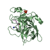
| ||||||||
| Unit cell |
|
- Components
| #1: Protein/peptide | Mass: 1157.368 Da / Num. of mol.: 2 / Source method: obtained synthetically / Source: (synth.) Phage display vector pTDisp (others) #2: Protein | Mass: 27554.158 Da / Num. of mol.: 2 / Mutation: C301A Source method: isolated from a genetically manipulated source Source: (gene. exp.)  Production host:  References: UniProt: P06869, u-plasminogen activator #3: Chemical | ChemComp-PO4 / #4: Water | ChemComp-HOH / | Has protein modification | Y | |
|---|
-Experimental details
-Experiment
| Experiment | Method:  X-RAY DIFFRACTION / Number of used crystals: 1 X-RAY DIFFRACTION / Number of used crystals: 1 |
|---|
- Sample preparation
| Crystal | Density Matthews: 3.37 Å3/Da / Density % sol: 63.55 % |
|---|---|
| Crystal grow | Temperature: 298.15 K / Method: vapor diffusion, sitting drop / pH: 8.5 Details: 80 mM Tris-HCl pH 8.5, 1.6 M NaH2PO4 and 20% Glycerol |
-Data collection
| Diffraction | Mean temperature: 100 K | ||||||||||||||||||||||||
|---|---|---|---|---|---|---|---|---|---|---|---|---|---|---|---|---|---|---|---|---|---|---|---|---|---|
| Diffraction source | Source:  SYNCHROTRON / Site: SYNCHROTRON / Site:  SSRF SSRF  / Beamline: BL19U1 / Wavelength: 0.979 Å / Beamline: BL19U1 / Wavelength: 0.979 Å | ||||||||||||||||||||||||
| Detector | Type: DECTRIS PILATUS3 S 6M / Detector: PIXEL / Date: Dec 17, 2017 | ||||||||||||||||||||||||
| Radiation | Protocol: SINGLE WAVELENGTH / Monochromatic (M) / Laue (L): M / Scattering type: x-ray | ||||||||||||||||||||||||
| Radiation wavelength | Wavelength: 0.979 Å / Relative weight: 1 | ||||||||||||||||||||||||
| Reflection | Resolution: 2.53→71.39 Å / Num. obs: 26295 / % possible obs: 100 % / Redundancy: 10 % / Biso Wilson estimate: 50.738 Å2 / Rpim(I) all: 0.037 / Rrim(I) all: 0.117 / Net I/σ(I): 16 / Num. measured all: 262704 | ||||||||||||||||||||||||
| Reflection shell | Diffraction-ID: 1
|
-Phasing
| Phasing | Method:  molecular replacement molecular replacement |
|---|
- Processing
| Software |
| ||||||||||||||||||||||||||||||||||||||||
|---|---|---|---|---|---|---|---|---|---|---|---|---|---|---|---|---|---|---|---|---|---|---|---|---|---|---|---|---|---|---|---|---|---|---|---|---|---|---|---|---|---|
| Refinement | Method to determine structure:  MOLECULAR REPLACEMENT MOLECULAR REPLACEMENTStarting model: 4DVA Resolution: 2.53→71.389 Å / SU ML: 0.38 / Cross valid method: THROUGHOUT / σ(F): 1.34 / Phase error: 29.41 / Stereochemistry target values: ML
| ||||||||||||||||||||||||||||||||||||||||
| Solvent computation | Shrinkage radii: 0.9 Å / VDW probe radii: 1.11 Å / Solvent model: FLAT BULK SOLVENT MODEL | ||||||||||||||||||||||||||||||||||||||||
| Displacement parameters | Biso max: 139.73 Å2 / Biso mean: 61.3391 Å2 / Biso min: 32.2 Å2 | ||||||||||||||||||||||||||||||||||||||||
| Refinement step | Cycle: final / Resolution: 2.53→71.389 Å
| ||||||||||||||||||||||||||||||||||||||||
| Refinement TLS params. | Method: refined / Origin x: 33.429 Å / Origin y: -34.866 Å / Origin z: 30.745 Å
| ||||||||||||||||||||||||||||||||||||||||
| Refinement TLS group | Selection details: all |
 Movie
Movie Controller




 PDBj
PDBj


