[English] 日本語
Yorodumi
- PDB-4ecm: 2.3 Angstrom Crystal Structure of a Glucose-1-phosphate Thymidyly... -

+
Open data


ID or keywords:

Loading...

-
Basic information

Entry
Database: PDB / ID: 4ecm
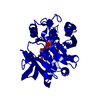
Title
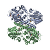
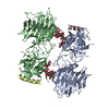
2.3 Angstrom Crystal Structure of a Glucose-1-phosphate Thymidylyltransferase from Bacillus anthracis in Complex with Thymidine-5-diphospho-alpha-D-glucose and Pyrophosphate
ComponentsGlucose-1-phosphate thymidylyltransferase
KeywordsTRANSFERASE / Glucose-1-phosphate Thymidylyltransferase / Structural Genomics / Center for Structural Genomics of Infectious Diseases / CSGID
Function / homology
Function and homology information


glucose-1-phosphate thymidylyltransferase / glucose-1-phosphate thymidylyltransferase activity / extracellular polysaccharide biosynthetic process
Similarity search - Function
Glucose-1-phosphate thymidylyltransferase, short form / Nucleotidyl transferase domain / Nucleotidyl transferase / Spore Coat Polysaccharide Biosynthesis Protein SpsA; Chain A / Spore Coat Polysaccharide Biosynthesis Protein SpsA; Chain A / Nucleotide-diphospho-sugar transferases / Alpha-Beta Complex / Alpha Beta
Similarity search - Domain/homology
2'DEOXY-THYMIDINE-5'-DIPHOSPHO-ALPHA-D-GLUCOSE / PYROPHOSPHATE 2- / glucose-1-phosphate thymidylyltransferase / glucose-1-phosphate thymidylyltransferase
Similarity search - Component
Biological speciesBacillus anthracis (anthrax bacterium)
MethodX-RAY DIFFRACTION / SYNCHROTRON / MOLECULAR REPLACEMENT / Resolution: 2.3 Å
AuthorsMinasov, G. / Kuhn, M. / Halavaty, A. / Shuvalova, L. / Dubrovska, I. / Winsor, J. / Papazisi, L. / Anderson, W.F. / Center for Structural Genomics of Infectious Diseases (CSGID)
CitationJournal: Acta Crystallogr F Struct Biol Commun / Year: 2017
Title: Structure of the Bacillus anthracis dTDP-L-rhamnose-biosynthetic enzyme glucose-1-phosphate thymidylyltransferase (RfbA).
Authors: Baumgartner, J. / Lee, J. / Halavaty, A.S. / Minasov, G. / Anderson, W.F. / Kuhn, M.L.
History
DepositionMar 26, 2012Deposition site: RCSB / Processing site: RCSB
Revision 1.0Apr 4, 2012Provider: repository / Type: Initial release
Revision 1.1May 23, 2012Group: Atomic model
Revision 1.2Nov 15, 2017Group: Database references / Refinement description / Category: citation / citation_author / software
Item: _citation.country / _citation.journal_abbrev ..._citation.country / _citation.journal_abbrev / _citation.journal_id_ASTM / _citation.journal_id_CSD / _citation.journal_id_ISSN / _citation.journal_volume / _citation.page_first / _citation.page_last / _citation.pdbx_database_id_DOI / _citation.pdbx_database_id_PubMed / _citation.title / _citation.year / _software.name
Revision 1.3Sep 13, 2023Group: Data collection / Database references ...Data collection / Database references / Derived calculations / Refinement description
Category: chem_comp_atom / chem_comp_bond ...chem_comp_atom / chem_comp_bond / database_2 / pdbx_initial_refinement_model / pdbx_struct_special_symmetry / struct_ref_seq_dif / struct_site
Item: _database_2.pdbx_DOI / _database_2.pdbx_database_accession ..._database_2.pdbx_DOI / _database_2.pdbx_database_accession / _struct_ref_seq_dif.details / _struct_site.pdbx_auth_asym_id / _struct_site.pdbx_auth_comp_id / _struct_site.pdbx_auth_seq_id

-
Structure visualization

Structure viewerMolecule:
MolmilJmol/JSmol

Downloads & links

-
Assembly

Deposited unit
A: Glucose-1-phosphate thymidylyltransferase
hetero molecules


Theoretical massNumber of molelcules
Total (without water)31,0193
Polymers30,2781
Non-polymers7402
Water4,071226
1
A: Glucose-1-phosphate thymidylyltransferase
hetero molecules

A: Glucose-1-phosphate thymidylyltransferase
hetero molecules

A: Glucose-1-phosphate thymidylyltransferase
hetero molecules

A: Glucose-1-phosphate thymidylyltransferase
hetero molecules


Theoretical massNumber of molelcules
Total (without water)124,07412
Polymers121,1134
Non-polymers2,9618
Water724
TypeNameSymmetry operationNumber
identity operation1_555x,y,z1
crystal symmetry operation4_655-x+1,-y,z1
crystal symmetry operation8_556x-y,-y,-z+11
crystal symmetry operation11_656-x+y+1,y,-z+11
Unit cell
Length a, b, c (Å)135.072, 135.072, 85.138
Angle α, β, γ (deg.)90.00, 90.00, 120.00
Int Tables number177
Space group name H-MP622
Components on special symmetry positions
IDModelComponents
11A--4-

PHE

-
Components

#1: Protein Glucose-1-phosphate thymidylyltransferase


Mass: 30278.312 Da / Num. of mol.: 1
Source method: isolated from a genetically manipulated source
Source: (gene. exp.) Bacillus anthracis (anthrax bacterium) / Gene: BA1228, BAS1135, BA_1228, GBAA_1228 / Plasmid: pMCSG7 / Production host: Escherichia coli (E. coli) / Strain (production host): BL21(DE3)
References: UniProt: Q81TP2, UniProt: A0A6L8PCC3*PLUS, glucose-1-phosphate thymidylyltransferase
#2: Chemical ChemComp-DAU / 2'DEOXY-THYMIDINE-5'-DIPHOSPHO-ALPHA-D-GLUCOSE


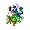
Mass: 564.329 Da / Num. of mol.: 1 / Source method: obtained synthetically / Formula: C16H26N2O16P2
#3: Chemical ChemComp-POP / PYROPHOSPHATE 2-


Mass: 175.959 Da / Num. of mol.: 1 / Source method: obtained synthetically / Formula: H2O7P2
#4: Water ChemComp-HOH / water


Mass: 18.015 Da / Num. of mol.: 226 / Source method: isolated from a natural source / Formula: H2O

-
Experimental details

-
Experiment

ExperimentMethod: X-RAY DIFFRACTION / Number of used crystals: 1

-
Sample preparation

CrystalDensity Matthews: 3.7 Å3/Da / Density % sol: 66.78 %
Crystal growTemperature: 295 K / Method: vapor diffusion, sitting drop / pH: 8.5
Details: Protein: 7.5 mg/mL, 0.5M NaCl, 0.01M Tris-HCl (pH 8.3), 0.001M Thymidine-5-diphospho-alpha-D-glucose. Screen: PACT (H8), 0.2 Sodium Sulfate, 0.1M TRIS propane, 20% (w/v) PEG 33500, VAPOR ...Details: Protein: 7.5 mg/mL, 0.5M NaCl, 0.01M Tris-HCl (pH 8.3), 0.001M Thymidine-5-diphospho-alpha-D-glucose. Screen: PACT (H8), 0.2 Sodium Sulfate, 0.1M TRIS propane, 20% (w/v) PEG 33500, VAPOR DIFFUSION, SITTING DROP, temperature 295K

-
Data collection

DiffractionMean temperature: 100 K
Diffraction sourceSource: SYNCHROTRON / Site: APS / Beamline: 21-ID-G / Wavelength: 0.97856 Å
DetectorType: MARMOSAIC 300 mm CCD / Detector: CCD / Date: Mar 5, 2012 / Details: Beryllium lenses
RadiationMonochromator: Diamond / Protocol: SINGLE WAVELENGTH / Monochromatic (M) / Laue (L): M / Scattering type: x-ray
Radiation wavelengthWavelength: 0.97856 Å / Relative weight: 1
ReflectionResolution: 2.3→30 Å / Num. all: 20938 / Num. obs: 20938 / % possible obs: 99.9 % / Observed criterion σ(I): -3 / Redundancy: 10.7 % / Biso Wilson estimate: 32.4 Å2 / Rmerge(I) obs: 0.108 / Net I/σ(I): 22.9
Reflection shellResolution: 2.3→2.34 Å / Redundancy: 10.9 % / Rmerge(I) obs: 0.533 / Mean I/σ(I) obs: 5.1 / Num. unique all: 1022 / % possible all: 100

-
Processing

Software
NameVersionClassification
Blu-IceMaxdata collection
PHASERphasing
REFMAC5.5.0102refinement
HKL-3000data reduction
HKL-3000data scaling
RefinementMethod to determine structure: MOLECULAR REPLACEMENT
Starting model: PDB entry 3HL3
Resolution: 2.3→29.24 Å / Cor.coef. Fo:Fc: 0.964 / Cor.coef. Fo:Fc free: 0.95 / SU B: 8.229 / SU ML: 0.094
Isotropic thermal model: Thermal Factors Individually Refined
Cross valid method: THROUGHOUT / ESU R: 0.171 / ESU R Free: 0.151 / Stereochemistry target values: MAXIMUM LIKELIHOOD / Details: HYDROGENS HAVE BEEN ADDED IN THE RIDING POSITIONS
RfactorNum. reflection% reflectionSelection details
Rfree0.18588 1073 5.1 %RANDOM
Rwork0.15271 ---
all0.15442 19775 --
obs0.15442 19775 99.92 %-
Solvent computationIon probe radii: 0.8 Å / Shrinkage radii: 0.8 Å / VDW probe radii: 1.2 Å / Solvent model: MASK
Displacement parametersBiso mean: 24.768 Å2
Baniso -1Baniso -2Baniso -3
1--0.26 Å2-0.13 Å20 Å2
2---0.26 Å20 Å2
3---0.39 Å2
Refinement stepCycle: LAST / Resolution: 2.3→29.24 Å
ProteinNucleic acidLigandSolventTotal
Num. atoms1940 0 45 226 2211
Refine LS restraints
Refine-IDTypeDev idealDev ideal targetNumber
X-RAY DIFFRACTIONr_bond_refined_d0.0110.0222187
X-RAY DIFFRACTIONr_bond_other_d0.0010.021478
X-RAY DIFFRACTIONr_angle_refined_deg1.5261.9762983
X-RAY DIFFRACTIONr_angle_other_deg0.8533613
X-RAY DIFFRACTIONr_dihedral_angle_1_deg3.5565276
X-RAY DIFFRACTIONr_dihedral_angle_2_deg31.56724.216102
X-RAY DIFFRACTIONr_dihedral_angle_3_deg10.42315379
X-RAY DIFFRACTIONr_dihedral_angle_4_deg19.9291514
X-RAY DIFFRACTIONr_chiral_restr0.1110.2321
X-RAY DIFFRACTIONr_gen_planes_refined0.0050.022480
X-RAY DIFFRACTIONr_gen_planes_other0.0020.02453
X-RAY DIFFRACTIONr_mcbond_it1.0441.51314
X-RAY DIFFRACTIONr_mcbond_other0.2851.5541
X-RAY DIFFRACTIONr_mcangle_it1.9322142
X-RAY DIFFRACTIONr_scbond_it3.0093873
X-RAY DIFFRACTIONr_scangle_it4.5514.5841
LS refinement shellResolution: 2.3→2.359 Å / Total num. of bins used: 20
RfactorNum. reflection% reflection
Rfree0.206 81 -
Rwork0.154 1416 -
obs-1416 100 %
Refinement TLS params.

Method: refined / Refine-ID: X-RAY DIFFRACTION

IDL112)L122)L132)L222)L232)L332)S11 (Å °)S12 (Å °)S13 (Å °)S21 (Å °)S22 (Å °)S23 (Å °)S31 (Å °)S32 (Å °)S33 (Å °)T112)T122)T132)T222)T232)T332)Origin x (Å)Origin y (Å)Origin z (Å)
111.896326.30897.198477.426631.167216.44961.01230.3263-0.195-0.5147-1.4531-0.1708-1.2656-1.77150.44080.50280.1628-0.02960.5828-0.29610.677367.7583-16.36490.3832
20.2385-0.2210.81490.9586-1.32923.2388-0.0034-0.0594-0.0113-0.03350.14660.1218-0.0222-0.2785-0.14320.09220.0215-0.00490.03260.01050.099360.6445-13.165529.9024
30.38680.14410.09340.7168-0.1640.77590.0117-0.00410.009-0.0389-0.0060.0389-0.00250.0543-0.00570.08450.01580.00130.0080.00270.053967.4003-13.025724.0596
40.5740.12570.25622.8831-0.46060.670.01210.0122-0.0434-0.02880.00560.13860.0703-0.1378-0.01770.095-0.0304-0.02420.0476-0.0130.053852.9618-28.565619.4155
52.73721.0588-0.1663.8413-0.92041.8757-0.0674-0.19840.32710.04340.06380.6212-0.2415-0.32350.00360.1240.0247-0.03340.0894-0.01090.154950.4555-13.570314.2576
60.8644-0.05890.5990.7816-0.01761.27550.0084-0.0114-0.015-0.02410.0133-0.09860.209-0.0031-0.02170.06920.00240.0050.002-0.00950.064968.2182-25.679429.428
Refinement TLS group
IDRefine-IDRefine TLS-IDAuth asym-IDAuth seq-ID
1X-RAY DIFFRACTION1A-4
2X-RAY DIFFRACTION2A0 - 16
3X-RAY DIFFRACTION3A17 - 130
4X-RAY DIFFRACTION4A131 - 175
5X-RAY DIFFRACTION5A176 - 206
6X-RAY DIFFRACTION6A207 - 243

+
About Yorodumi

-
News

-
Feb 9, 2022. New format data for meta-information of EMDB entries

New format data for meta-information of EMDB entries

  • Version 3 of the EMDB header file is now the official format.
  • The previous official version 1.9 will be removed from the archive.

Related info.:EMDB header

External links:wwPDB to switch to version 3 of the EMDB data model

-
Aug 12, 2020. Covid-19 info

Covid-19 info

URL: https://pdbjlvh1.pdbj.org/emnavi/covid19.php

New page: Covid-19 featured information page in EM Navigator.

Related info.:Covid-19 info / Mar 5, 2020. Novel coronavirus structure data

+
Mar 5, 2020. Novel coronavirus structure data

Novel coronavirus structure data

Related info.:Yorodumi Speices / Aug 12, 2020. Covid-19 info

External links:COVID-19 featured content - PDBj / Molecule of the Month (242):Coronavirus Proteases

+
Jan 31, 2019. EMDB accession codes are about to change! (news from PDBe EMDB page)

EMDB accession codes are about to change! (news from PDBe EMDB page)

  • The allocation of 4 digits for EMDB accession codes will soon come to an end. Whilst these codes will remain in use, new EMDB accession codes will include an additional digit and will expand incrementally as the available range of codes is exhausted. The current 4-digit format prefixed with “EMD-” (i.e. EMD-XXXX) will advance to a 5-digit format (i.e. EMD-XXXXX), and so on. It is currently estimated that the 4-digit codes will be depleted around Spring 2019, at which point the 5-digit format will come into force.
  • The EM Navigator/Yorodumi systems omit the EMD- prefix.

Related info.:Q: What is EMD? / ID/Accession-code notation in Yorodumi/EM Navigator

External links:EMDB Accession Codes are Changing Soon! / Contact to PDBj

+
Jul 12, 2017. Major update of PDB

Major update of PDB

  • wwPDB released updated PDB data conforming to the new PDBx/mmCIF dictionary.
  • This is a major update changing the version number from 4 to 5, and with Remediation, in which all the entries are updated.
  • In this update, many items about electron microscopy experimental information are reorganized (e.g. em_software).
  • Now, EM Navigator and Yorodumi are based on the updated data.

External links:wwPDB Remediation / Enriched Model Files Conforming to OneDep Data Standards Now Available in the PDB FTP Archive

-
Yorodumi

Thousand views of thousand structures

  • Yorodumi is a browser for structure data from EMDB, PDB, SASBDB, etc.
  • This page is also the successor to EM Navigator detail page, and also detail information page/front-end page for Omokage search.
  • The word "yorodu" (or yorozu) is an old Japanese word meaning "ten thousand". "mi" (miru) is to see.

Related info.:EMDB / PDB / SASBDB / Comparison of 3 databanks / Yorodumi Search / Aug 31, 2016. New EM Navigator & Yorodumi / Yorodumi Papers / Jmol/JSmol / Function and homology information / Changes in new EM Navigator and Yorodumi

Read more