 Movie
Movie Controller


+ Open data

- Basic information
| Entry | Database: PDB / ID: 6a6y | ||||||
|---|---|---|---|---|---|---|---|
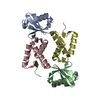
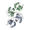
| Title | Crystal Structure of Asf1 from Plasmodium falciparum | ||||||
 Components Components | Histone chaperone ASF1, putative | ||||||
 Keywords Keywords | CHAPERONE / Histone chaperone / Chromatin assembly and disassembly / Plasmodium facliparum | ||||||
| Function / homology |  Function and homology information Function and homology informationDNA replication-dependent chromatin assembly / nucleosome disassembly / : / nucleosome assembly / histone binding / chromatin binding / chromatin / nucleus Similarity search - Function | ||||||
| Biological species |  | ||||||
| Method |  X-RAY DIFFRACTION / MOLECULAR REPLACEMENT / Resolution: 2.491 Å X-RAY DIFFRACTION / MOLECULAR REPLACEMENT / Resolution: 2.491 Å | ||||||
 Authors Authors | Srivastava, D.K. / Roy, S. | ||||||
 Citation Citation | Journal: To Be Published Title: Structural and Functional characterization of As1 from Plasmodium falciparum Authors: Srivastava, D.K. / Gunjan, S. / Seshadri, V. / Roy, S. | ||||||
| History |
|
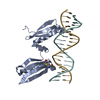
- Structure visualization
| Structure viewer | Molecule: MolmilJmol/JSmol |
|---|
- Downloads & links
-Download
| PDBx/mmCIF format | 6a6y.cif.gz | 141.8 KB | Display | PDBx/mmCIF format |
|---|---|---|---|---|
| PDB format | pdb6a6y.ent.gz | 111.1 KB | Display | PDB format |
| PDBx/mmJSON format | 6a6y.json.gz | Tree view | PDBx/mmJSON format | |
| Others |  Other downloads Other downloads |
-Validation report
| Arichive directory | https://data.pdbj.org/pub/pdb/validation_reports/a6/6a6yftp://data.pdbj.org/pub/pdb/validation_reports/a6/6a6y | HTTPS FTP |
|---|
-Related structure data
| Related structure data | 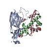 2hueS S: Starting model for refinement |
|---|---|
| Similar structure data |


-Links
 PDBj
PDBj

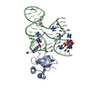
- Assembly
| Deposited unit | 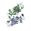
| ||||||||
|---|---|---|---|---|---|---|---|---|---|
| 1 |
| ||||||||
| Unit cell |
|
-Components
| #1: Protein | Mass: 18714.309 Da / Num. of mol.: 2 Source method: isolated from a genetically manipulated source Source: (gene. exp.) Gene: PF3D7_1224500 / Production host:  #2: Chemical | ChemComp-SCN /   Mass: 58.082 Da / Num. of mol.: 6 / Source method: obtained synthetically / Formula: CNS Mass: 58.082 Da / Num. of mol.: 6 / Source method: obtained synthetically / Formula: CNS#3: Chemical |   Mass: 62.068 Da / Num. of mol.: 2 / Source method: obtained synthetically / Formula: C2H6O2 Mass: 62.068 Da / Num. of mol.: 2 / Source method: obtained synthetically / Formula: C2H6O2#4: Water | ChemComp-HOH / |  Mass: 18.015 Da / Num. of mol.: 129 / Source method: isolated from a natural source / Formula: H2O Mass: 18.015 Da / Num. of mol.: 129 / Source method: isolated from a natural source / Formula: H2O |
|---|
-Experimental details
-Experiment
| Experiment | Method: X-RAY DIFFRACTION / Number of used crystals: 1 |
|---|
- Sample preparation
| Crystal | Density Matthews: 2.68 Å3/Da / Density % sol: 54.1 % |
|---|---|
| Crystal grow | Temperature: 293 K / Method: vapor diffusion, hanging drop / pH: 6.5 Details: 30% PEG 3350, 0.2M Sodium thiocyanate, 0.1M MES pH 6.5 |
-Data collection
| Diffraction | Mean temperature: 100 K |
|---|---|
| Diffraction source | Source: ROTATING ANODE / Type: RIGAKU ULTRAX 18 / Wavelength: 1.5418 Å |
| Detector | Type: RIGAKU RAXIS IV++ / Detector: IMAGE PLATE / Date: Sep 25, 2014 / Details: mirror |
| Radiation | Protocol: SINGLE WAVELENGTH / Monochromatic (M) / Laue (L): M / Scattering type: x-ray |
| Radiation wavelength | Wavelength: 1.5418 Å / Relative weight: 1 |
| Reflection | Resolution: 2.491→44.36 Å / Num. obs: 13132 / % possible obs: 97.8 % / Redundancy: 7.5 % / Biso Wilson estimate: 41.79 Å2 / CC1/2: 0.997 / Rmerge(I) obs: 0.1232 / Rpim(I) all: 0.04838 / Rrim(I) all: 0.1324 / Net I/σ(I): 13.86 |
| Reflection shell | Resolution: 2.491→2.58 Å / Redundancy: 7.3 % / Num. unique obs: 1258 / CC1/2: 0.831 / % possible all: 95.66 |
- Processing
| Software |
| |||||||||||||||||||||||||||||||||||||||||||||||||||||||||||||||||||||||||||
|---|---|---|---|---|---|---|---|---|---|---|---|---|---|---|---|---|---|---|---|---|---|---|---|---|---|---|---|---|---|---|---|---|---|---|---|---|---|---|---|---|---|---|---|---|---|---|---|---|---|---|---|---|---|---|---|---|---|---|---|---|---|---|---|---|---|---|---|---|---|---|---|---|---|---|---|---|
| Refinement | Method to determine structure: MOLECULAR REPLACEMENT Starting model: 2HUE Resolution: 2.491→44.359 Å / SU ML: 0.32 / Cross valid method: FREE R-VALUE / σ(F): 1.35 / Phase error: 24.76
| |||||||||||||||||||||||||||||||||||||||||||||||||||||||||||||||||||||||||||
| Solvent computation | Shrinkage radii: 0.9 Å / VDW probe radii: 1.11 Å | |||||||||||||||||||||||||||||||||||||||||||||||||||||||||||||||||||||||||||
| Refinement step | Cycle: LAST / Resolution: 2.491→44.359 Å
| |||||||||||||||||||||||||||||||||||||||||||||||||||||||||||||||||||||||||||
| Refine LS restraints |
| |||||||||||||||||||||||||||||||||||||||||||||||||||||||||||||||||||||||||||
| LS refinement shell |
| |||||||||||||||||||||||||||||||||||||||||||||||||||||||||||||||||||||||||||
| Refinement TLS params. | Method: refined / Refine-ID: X-RAY DIFFRACTION
| |||||||||||||||||||||||||||||||||||||||||||||||||||||||||||||||||||||||||||
| Refinement TLS group |
|
