 Movie
Movie Controller

[English] 日本語
 Yorodumi
- PDB-6a6t: Crystal structure of the modified fructosyl peptide oxidase from ... -
+ Open data

- Basic information
| Entry | Database: PDB / ID: 6a6t | ||||||
|---|---|---|---|---|---|---|---|
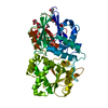
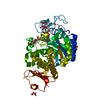
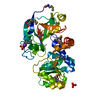
| Title | Crystal structure of the modified fructosyl peptide oxidase from Aspergillus nidulans with R61G mutation | ||||||
 Components Components | Fructosyl amine: oxygen oxidoreductase | ||||||
 Keywords Keywords | OXIDOREDUCTASE / fructosyl peptide / Aspergillus nidulans / FAD-binding protein | ||||||
| Function / homology | D-Amino Acid Oxidase, subunit A, domain 2 / D-Amino Acid Oxidase; Chain A, domain 2 / FAD/NAD(P)-binding domain / FAD/NAD(P)-binding domain / 3-Layer(bba) Sandwich / 2-Layer Sandwich / Alpha Beta / (4S,5S)-1,2-DITHIANE-4,5-DIOL / FLAVIN-ADENINE DINUCLEOTIDE Function and homology information Function and homology information | ||||||
| Biological species |  | ||||||
| Method |  X-RAY DIFFRACTION / SYNCHROTRON / MOLECULAR REPLACEMENT / Resolution: 1.901 Å X-RAY DIFFRACTION / SYNCHROTRON / MOLECULAR REPLACEMENT / Resolution: 1.901 Å | ||||||
 Authors Authors | Ogawa, N. / Maruyama, Y. / Itoh, T. / Hashimoto, W. / Murata, K. | ||||||
 Citation Citation | Journal: Sci Rep / Year: 2019 Title: Creation of haemoglobin A1c direct oxidase from fructosyl peptide oxidase by combined structure-based site specific mutagenesis and random mutagenesis. Authors: Ogawa, N. / Kimura, T. / Umehara, F. / Katayama, Y. / Nagai, G. / Suzuki, K. / Aisaka, K. / Maruyama, Y. / Itoh, T. / Hashimoto, W. / Murata, K. / Ichimura, M. | ||||||
| History |
|
- Structure visualization
| Structure viewer | Molecule: MolmilJmol/JSmol |
|---|
- Downloads & links
-Download
| PDBx/mmCIF format | 6a6t.cif.gz | 189.5 KB | Display | PDBx/mmCIF format |
|---|---|---|---|---|
| PDB format | pdb6a6t.ent.gz | 149.3 KB | Display | PDB format |
| PDBx/mmJSON format | 6a6t.json.gz | Tree view | PDBx/mmJSON format | |
| Others |  Other downloads Other downloads |
-Validation report
| Summary document | 6a6t_validation.pdf.gz | 747.2 KB | Display | wwPDB validaton report |
|---|---|---|---|---|
| Full document | 6a6t_full_validation.pdf.gz | 753.5 KB | Display | |
| Data in XML | 6a6t_validation.xml.gz | 19.6 KB | Display | |
| Data in CIF | 6a6t_validation.cif.gz | 27.7 KB | Display | |
| Arichive directory | https://data.pdbj.org/pub/pdb/validation_reports/a6/6a6tftp://data.pdbj.org/pub/pdb/validation_reports/a6/6a6t | HTTPS FTP |
-Related structure data
| Related structure data |  6a6rSC  6a6sC  6a6uC  6a6vC S: Starting model for refinement C: citing same article ( |
|---|---|
| Similar structure data |
-Links
 PDBj
PDBj- Assembly
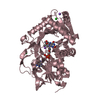
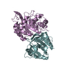
| Deposited unit | 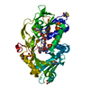
| ||||||||
|---|---|---|---|---|---|---|---|---|---|
| 1 |
| ||||||||
| Unit cell |
|
-Components
| #1: Protein | Mass: 48621.438 Da / Num. of mol.: 1 Source method: isolated from a genetically manipulated source Source: (gene. exp.)  | ||||||
|---|---|---|---|---|---|---|---|
| #2: Chemical | ChemComp-FAD /   Mass: 785.550 Da / Num. of mol.: 1 / Source method: obtained synthetically / Formula: C27H33N9O15P2 / Comment: FAD*YM Mass: 785.550 Da / Num. of mol.: 1 / Source method: obtained synthetically / Formula: C27H33N9O15P2 / Comment: FAD*YM | ||||||
| #3: Chemical | ChemComp-SO4 /   Mass: 96.063 Da / Num. of mol.: 4 / Source method: obtained synthetically / Formula: SO4 Mass: 96.063 Da / Num. of mol.: 4 / Source method: obtained synthetically / Formula: SO4#4: Chemical | ChemComp-D1D / (   Mass: 152.235 Da / Num. of mol.: 5 / Source method: obtained synthetically / Formula: C4H8O2S2 Mass: 152.235 Da / Num. of mol.: 5 / Source method: obtained synthetically / Formula: C4H8O2S2#5: Water | ChemComp-HOH / |  Mass: 18.015 Da / Num. of mol.: 159 / Source method: isolated from a natural source / Formula: H2O Mass: 18.015 Da / Num. of mol.: 159 / Source method: isolated from a natural source / Formula: H2OHas protein modification | Y | |
-Experimental details
-Experiment
| Experiment | Method: X-RAY DIFFRACTION / Number of used crystals: 1 |
|---|
- Sample preparation
| Crystal | Density Matthews: 2.49 Å3/Da / Density % sol: 50.56 % |
|---|---|
| Crystal grow | Temperature: 293 K / Method: vapor diffusion, hanging drop / pH: 10.4 Details: 1.75 M ammonium sulfate, 0.2 M lithium sulfate, and 0.08 M CAPS (N-cyclohexyl-3-aminopropanesulfonic acid) buffer (pH 10.4) |
-Data collection
| Diffraction | Mean temperature: 100 K |
|---|---|
| Diffraction source | Source: SYNCHROTRON / Site: SPring-8  / Beamline: BL38B1 / Wavelength: 1 Å / Beamline: BL38B1 / Wavelength: 1 Å |
| Detector | Type: ADSC QUANTUM 210 / Detector: CCD / Date: Dec 9, 2011 |
| Radiation | Protocol: SINGLE WAVELENGTH / Monochromatic (M) / Laue (L): M / Scattering type: x-ray |
| Radiation wavelength | Wavelength: 1 Å / Relative weight: 1 |
| Reflection | Resolution: 1.9→50 Å / Num. obs: 38889 / % possible obs: 99.6 % / Redundancy: 2.6 % / Rmerge(I) obs: 0.057 / Net I/σ(I): 14.1 |
| Reflection shell | Resolution: 1.9→1.93 Å / Rmerge(I) obs: 0.405 / Num. unique obs: 1893 |
- Processing
| Software |
| ||||||||||||||||||||||||||||||||||||||||||||||||||||||||||||||||||||||||||||||||||||||||||||||||||||
|---|---|---|---|---|---|---|---|---|---|---|---|---|---|---|---|---|---|---|---|---|---|---|---|---|---|---|---|---|---|---|---|---|---|---|---|---|---|---|---|---|---|---|---|---|---|---|---|---|---|---|---|---|---|---|---|---|---|---|---|---|---|---|---|---|---|---|---|---|---|---|---|---|---|---|---|---|---|---|---|---|---|---|---|---|---|---|---|---|---|---|---|---|---|---|---|---|---|---|---|---|---|
| Refinement | Method to determine structure: MOLECULAR REPLACEMENT Starting model: 6A6R Resolution: 1.901→33.732 Å / SU ML: 0.26 / Cross valid method: FREE R-VALUE / σ(F): 1.35 / Phase error: 31.76
| ||||||||||||||||||||||||||||||||||||||||||||||||||||||||||||||||||||||||||||||||||||||||||||||||||||
| Solvent computation | Shrinkage radii: 0.9 Å / VDW probe radii: 1.11 Å | ||||||||||||||||||||||||||||||||||||||||||||||||||||||||||||||||||||||||||||||||||||||||||||||||||||
| Refinement step | Cycle: LAST / Resolution: 1.901→33.732 Å
| ||||||||||||||||||||||||||||||||||||||||||||||||||||||||||||||||||||||||||||||||||||||||||||||||||||
| Refine LS restraints |
| ||||||||||||||||||||||||||||||||||||||||||||||||||||||||||||||||||||||||||||||||||||||||||||||||||||
| LS refinement shell |
| ||||||||||||||||||||||||||||||||||||||||||||||||||||||||||||||||||||||||||||||||||||||||||||||||||||
| Refinement TLS params. | Method: refined / Refine-ID: X-RAY DIFFRACTION
| ||||||||||||||||||||||||||||||||||||||||||||||||||||||||||||||||||||||||||||||||||||||||||||||||||||
| Refinement TLS group |
|
