 Movie
Movie Controller

[English] 日本語
 Yorodumi
- PDB-5zni: Plasmodium falciparum purine nucleoside phosphorylase in complex ... -
+ Open data

- Basic information
| Entry | Database: PDB / ID: 5zni | ||||||
|---|---|---|---|---|---|---|---|
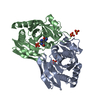
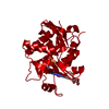
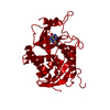
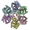
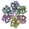
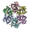
| Title | Plasmodium falciparum purine nucleoside phosphorylase in complex with mefloquine | ||||||
 Components Components | Purine nucleoside phosphorylase | ||||||
 Keywords Keywords | TRANSFERASE / nucleoside phosphorylase | ||||||
| Function / homology |  Function and homology information Function and homology informationS-methyl-5'-thioinosine phosphorylase / uridine catabolic process / guanosine phosphorylase activity / uridine phosphorylase activity / purine-nucleoside phosphorylase / purine-nucleoside phosphorylase activity / purine ribonucleoside salvage / cytosol Similarity search - Function | ||||||
| Biological species |  | ||||||
| Method |  X-RAY DIFFRACTION / SYNCHROTRON / MOLECULAR REPLACEMENT / Resolution: 2.3 Å X-RAY DIFFRACTION / SYNCHROTRON / MOLECULAR REPLACEMENT / Resolution: 2.3 Å | ||||||
 Authors Authors | Chen, D. / Nordlund, P. / Dziekan, J.M. | ||||||
 Citation Citation | Journal: Sci Transl Med / Year: 2019 Title: Identifying purine nucleoside phosphorylase as the target of quinine using cellular thermal shift assay. Authors: Dziekan, J.M. / Yu, H. / Chen, D. / Dai, L. / Wirjanata, G. / Larsson, A. / Prabhu, N. / Sobota, R.M. / Bozdech, Z. / Nordlund, P. | ||||||
| History |
|
- Structure visualization
| Structure viewer | Molecule: MolmilJmol/JSmol |
|---|
- Downloads & links
-Download
| PDBx/mmCIF format | 5zni.cif.gz | 61.3 KB | Display | PDBx/mmCIF format |
|---|---|---|---|---|
| PDB format | pdb5zni.ent.gz | 43.4 KB | Display | PDB format |
| PDBx/mmJSON format | 5zni.json.gz | Tree view | PDBx/mmJSON format | |
| Others |  Other downloads Other downloads |
-Validation report
| Arichive directory | https://data.pdbj.org/pub/pdb/validation_reports/zn/5zniftp://data.pdbj.org/pub/pdb/validation_reports/zn/5zni | HTTPS FTP |
|---|
-Related structure data
| Related structure data |  5zncC  1sq6S S: Starting model for refinement C: citing same article ( |
|---|---|
| Similar structure data |


-Links
 PDBj
PDBj- Assembly
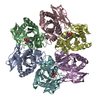
| Deposited unit | 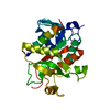
| ||||||||
|---|---|---|---|---|---|---|---|---|---|
| 1 | x 6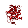
| ||||||||
| Unit cell |
|
-Components
| #1: Protein | Mass: 26891.156 Da / Num. of mol.: 1 Source method: isolated from a genetically manipulated source Source: (gene. exp.) Gene: PNP / Production host:  References: UniProt: Q8T9Z7, purine-nucleoside phosphorylase |
|---|---|
| #2: Chemical | ChemComp-PO4 /   Mass: 94.971 Da / Num. of mol.: 1 / Source method: obtained synthetically / Formula: PO4 Mass: 94.971 Da / Num. of mol.: 1 / Source method: obtained synthetically / Formula: PO4 |
| #3: Chemical | ChemComp-YMZ / (  Mass: 378.312 Da / Num. of mol.: 1 / Source method: obtained synthetically / Formula: C17H16F6N2O / Feature type: SUBJECT OF INVESTIGATION Mass: 378.312 Da / Num. of mol.: 1 / Source method: obtained synthetically / Formula: C17H16F6N2O / Feature type: SUBJECT OF INVESTIGATION |
| #4: Water | ChemComp-HOH /  Mass: 18.015 Da / Num. of mol.: 82 / Source method: isolated from a natural source / Formula: H2O Mass: 18.015 Da / Num. of mol.: 82 / Source method: isolated from a natural source / Formula: H2O |
| Has protein modification | N |
-Experimental details
-Experiment
| Experiment | Method: X-RAY DIFFRACTION / Number of used crystals: 1 |
|---|
- Sample preparation
| Crystal | Density Matthews: 2.23 Å3/Da / Density % sol: 44.91 % |
|---|---|
| Crystal grow | Temperature: 293 K / Method: vapor diffusion, sitting drop / Details: 0.2M magnesium formate |
-Data collection
| Diffraction | Mean temperature: 100 K | ||||||||||||||||||||||||||||||
|---|---|---|---|---|---|---|---|---|---|---|---|---|---|---|---|---|---|---|---|---|---|---|---|---|---|---|---|---|---|---|---|
| Diffraction source | Source: SYNCHROTRON / Site: Australian Synchrotron  / Beamline: MX1 / Wavelength: 1 Å / Beamline: MX1 / Wavelength: 1 Å | ||||||||||||||||||||||||||||||
| Detector | Type: ADSC QUANTUM 210r / Detector: CCD / Date: Jul 21, 2017 | ||||||||||||||||||||||||||||||
| Radiation | Protocol: SINGLE WAVELENGTH / Monochromatic (M) / Laue (L): M / Scattering type: x-ray | ||||||||||||||||||||||||||||||
| Radiation wavelength | Wavelength: 1 Å / Relative weight: 1 | ||||||||||||||||||||||||||||||
| Reflection | Resolution: 2.3→25.94 Å / Num. obs: 10842 / % possible obs: 99.6 % / Redundancy: 5.5 % / CC1/2: 0.998 / Rmerge(I) obs: 0.049 / Rpim(I) all: 0.023 / Rrim(I) all: 0.054 / Net I/σ(I): 21.6 / Num. measured all: 59291 / Scaling rejects: 330 | ||||||||||||||||||||||||||||||
| Reflection shell | Diffraction-ID: 1
|
- Processing
| Software |
| ||||||||||||||||||||||||||||||||||||||||||||||||||||||||||||
|---|---|---|---|---|---|---|---|---|---|---|---|---|---|---|---|---|---|---|---|---|---|---|---|---|---|---|---|---|---|---|---|---|---|---|---|---|---|---|---|---|---|---|---|---|---|---|---|---|---|---|---|---|---|---|---|---|---|---|---|---|---|
| Refinement | Method to determine structure: MOLECULAR REPLACEMENT Starting model: 1SQ6 Resolution: 2.3→25.94 Å / Cor.coef. Fo:Fc: 0.95 / Cor.coef. Fo:Fc free: 0.914 / SU B: 7.093 / SU ML: 0.172 / Cross valid method: THROUGHOUT / σ(F): 0 / ESU R: 0.349 / ESU R Free: 0.247 Details: HYDROGENS HAVE BEEN ADDED IN THE RIDING POSITIONS U VALUES : REFINED INDIVIDUALLY
| ||||||||||||||||||||||||||||||||||||||||||||||||||||||||||||
| Solvent computation | Ion probe radii: 0.8 Å / Shrinkage radii: 0.8 Å / VDW probe radii: 1.2 Å | ||||||||||||||||||||||||||||||||||||||||||||||||||||||||||||
| Displacement parameters | Biso max: 97.26 Å2 / Biso mean: 27.76 Å2 / Biso min: 6.81 Å2
| ||||||||||||||||||||||||||||||||||||||||||||||||||||||||||||
| Refinement step | Cycle: final / Resolution: 2.3→25.94 Å
| ||||||||||||||||||||||||||||||||||||||||||||||||||||||||||||
| Refine LS restraints |
| ||||||||||||||||||||||||||||||||||||||||||||||||||||||||||||
| LS refinement shell | Resolution: 2.3→2.36 Å / Rfactor Rfree error: 0 / Total num. of bins used: 20
|
