 Movie
Movie Controller

[English] 日本語
 Yorodumi
- PDB-5zmi: Crystal structure of APRT from Y. pseudotuberculosis in complex w... -
+ Open data

- Basic information
| Entry | Database: PDB / ID: 5zmi | ||||||
|---|---|---|---|---|---|---|---|
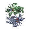
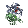
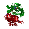
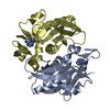
| Title | Crystal structure of APRT from Y. pseudotuberculosis in complex with adenine. | ||||||
 Components Components | Adenine phosphoribosyltransferase | ||||||
 Keywords Keywords | TRANSFERASE / adenine phosphoribosyltransferase / adenine | ||||||
| Function / homology |  Function and homology information Function and homology informationadenine salvage / adenine phosphoribosyltransferase / adenine phosphoribosyltransferase activity / AMP salvage / purine ribonucleoside salvage / cytosol Similarity search - Function | ||||||
| Biological species |  Yersinia pseudotuberculosis IP 32953 (bacteria) Yersinia pseudotuberculosis IP 32953 (bacteria) | ||||||
| Method |  X-RAY DIFFRACTION / SYNCHROTRON / MOLECULAR REPLACEMENT / Resolution: 2.05 Å X-RAY DIFFRACTION / SYNCHROTRON / MOLECULAR REPLACEMENT / Resolution: 2.05 Å | ||||||
 Authors Authors | Pavithra, G.C. / Fox, G.C. / Ramagopal, U.A. | ||||||
 Citation Citation | Journal: To be published Title: Crystal structure of adenine phosphoribosyltransferase from Yersinia pseudotuberculosis Authors: Pavithra, G.C. / Ramagopal, U.A. | ||||||
| History |
|
- Structure visualization
| Structure viewer | Molecule: MolmilJmol/JSmol |
|---|
- Downloads & links
-Download
| PDBx/mmCIF format | 5zmi.cif.gz | 83.6 KB | Display | PDBx/mmCIF format |
|---|---|---|---|---|
| PDB format | pdb5zmi.ent.gz | 61.5 KB | Display | PDB format |
| PDBx/mmJSON format | 5zmi.json.gz | Tree view | PDBx/mmJSON format | |
| Others |  Other downloads Other downloads |
-Validation report
| Arichive directory | https://data.pdbj.org/pub/pdb/validation_reports/zm/5zmiftp://data.pdbj.org/pub/pdb/validation_reports/zm/5zmi | HTTPS FTP |
|---|
-Related structure data
| Related structure data |  4mb6SC 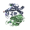 5zc7C S: Starting model for refinement C: citing same article ( |
|---|---|
| Similar structure data |


-Links
 PDBj
PDBj


- Assembly
| Deposited unit | 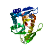
| ||||||||
|---|---|---|---|---|---|---|---|---|---|
| 1 | 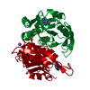
| ||||||||
| Unit cell |
|
-Components
| #1: Protein | Mass: 20165.990 Da / Num. of mol.: 1 Source method: isolated from a genetically manipulated source Source: (gene. exp.) Yersinia pseudotuberculosis IP 32953 (bacteria)Strain: IP32953 / Gene: YPTB0991 / Plasmid: pNIC28-Bsa4 / Production host: References: UniProt: Q66DQ2, adenine phosphoribosyltransferase |
|---|---|
| #2: Chemical | ChemComp-ADE /   Mass: 135.127 Da / Num. of mol.: 1 / Source method: obtained synthetically / Formula: C5H5N5 / Feature type: SUBJECT OF INVESTIGATION Mass: 135.127 Da / Num. of mol.: 1 / Source method: obtained synthetically / Formula: C5H5N5 / Feature type: SUBJECT OF INVESTIGATION |
| #3: Chemical | ChemComp-NA /   Mass: 22.990 Da / Num. of mol.: 1 / Source method: obtained synthetically / Formula: Na Mass: 22.990 Da / Num. of mol.: 1 / Source method: obtained synthetically / Formula: Na |
| #4: Water | ChemComp-HOH /  Mass: 18.015 Da / Num. of mol.: 10 / Source method: isolated from a natural source / Formula: H2O Mass: 18.015 Da / Num. of mol.: 10 / Source method: isolated from a natural source / Formula: H2O |
-Experimental details
-Experiment
| Experiment | Method: X-RAY DIFFRACTION / Number of used crystals: 1 |
|---|
- Sample preparation
| Crystal | Density Matthews: 2.79 Å3/Da / Density % sol: 55.95 % / Mosaicity: 0.68 ° |
|---|---|
| Crystal grow | Temperature: 298 K / Method: vapor diffusion, sitting drop / pH: 8.5 Details: 30% PEG4000, 0.1M Tris-Hcl pH 8.5, 0.2M Sodium Acetate with 5mM adenine and 5% ethylene glycol |
-Data collection
| Diffraction | Mean temperature: 100 K |
|---|---|
| Diffraction source | Source: SYNCHROTRON / Site: SOLEIL  / Beamline: PROXIMA 2 / Wavelength: 0.9677 Å / Beamline: PROXIMA 2 / Wavelength: 0.9677 Å |
| Detector | Type: DECTRIS EIGER X 9M / Detector: PIXEL / Date: Sep 29, 2017 |
| Radiation | Protocol: SINGLE WAVELENGTH / Monochromatic (M) / Laue (L): M / Scattering type: x-ray |
| Radiation wavelength | Wavelength: 0.9677 Å / Relative weight: 1 |
| Reflection | Resolution: 2.05→48.57 Å / Num. obs: 13665 / % possible obs: 98.1 % / Redundancy: 2.9 % / CC1/2: 0.996 / Rmerge(I) obs: 0.05 / Rpim(I) all: 0.034 / Rrim(I) all: 0.061 / Net I/σ(I): 9.9 |
| Reflection shell | Resolution: 2.05→2.11 Å / Redundancy: 2.4 % / Rmerge(I) obs: 0.49 / Num. unique obs: 1026 / CC1/2: 0.731 / Rpim(I) all: 0.375 / Rrim(I) all: 0.62 / % possible all: 94.8 |
- Processing
| Software |
| ||||||||||||||||||||||||||||||||||||||||||||||||||||||||||||
|---|---|---|---|---|---|---|---|---|---|---|---|---|---|---|---|---|---|---|---|---|---|---|---|---|---|---|---|---|---|---|---|---|---|---|---|---|---|---|---|---|---|---|---|---|---|---|---|---|---|---|---|---|---|---|---|---|---|---|---|---|---|
| Refinement | Method to determine structure: MOLECULAR REPLACEMENT Starting model: 4MB6 Resolution: 2.05→48.57 Å / Cor.coef. Fo:Fc: 0.972 / Cor.coef. Fo:Fc free: 0.957 / SU B: 11.233 / SU ML: 0.136 / SU R Cruickshank DPI: 0.1654 / Cross valid method: THROUGHOUT / σ(F): 0 / ESU R: 0.165 / ESU R Free: 0.155 Details: HYDROGENS HAVE BEEN ADDED IN THE RIDING POSITIONS U VALUES : WITH TLS ADDED
| ||||||||||||||||||||||||||||||||||||||||||||||||||||||||||||
| Solvent computation | Ion probe radii: 0.8 Å / Shrinkage radii: 0.8 Å / VDW probe radii: 1.2 Å | ||||||||||||||||||||||||||||||||||||||||||||||||||||||||||||
| Displacement parameters | Biso max: 102.09 Å2 / Biso mean: 58.491 Å2 / Biso min: 36.35 Å2
| ||||||||||||||||||||||||||||||||||||||||||||||||||||||||||||
| Refinement step | Cycle: final / Resolution: 2.05→48.57 Å
| ||||||||||||||||||||||||||||||||||||||||||||||||||||||||||||
| Refine LS restraints |
| ||||||||||||||||||||||||||||||||||||||||||||||||||||||||||||
| LS refinement shell | Resolution: 2.05→2.103 Å / Rfactor Rfree error: 0 / Total num. of bins used: 20
| ||||||||||||||||||||||||||||||||||||||||||||||||||||||||||||
| Refinement TLS params. | Method: refined / Origin x: 21.0714 Å / Origin y: -0.1791 Å / Origin z: 7.3287 Å
|
