 Movie
Movie Controller


+ Open data

- Basic information
| Entry | Database: PDB / ID: 2dy0 | ||||||
|---|---|---|---|---|---|---|---|
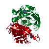
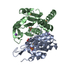
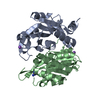
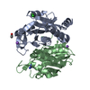
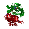
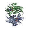
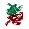
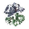
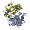
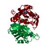
| Title | Crystal structure of project JW0458 from Escherichia coli | ||||||
 Components Components | Adenine phosphoribosyltransferase | ||||||
 Keywords Keywords | TRANSFERASE / structural genomics / NPPSFA / National Project on Protein Structural and Functional Analyses / RIKEN Structural Genomics/Proteomics Initiative / RSGI | ||||||
| Function / homology |  Function and homology information Function and homology informationadenine salvage / adenine phosphoribosyltransferase / adenine phosphoribosyltransferase activity / AMP salvage / purine ribonucleoside salvage / magnesium ion binding / protein homodimerization activity / cytosol Similarity search - Function | ||||||
| Biological species |  | ||||||
| Method |  X-RAY DIFFRACTION / SYNCHROTRON / MOLECULAR REPLACEMENT / Resolution: 1.25 Å X-RAY DIFFRACTION / SYNCHROTRON / MOLECULAR REPLACEMENT / Resolution: 1.25 Å | ||||||
 Authors Authors | Shimizu, K. / RIKEN Structural Genomics/Proteomics Initiative (RSGI) | ||||||
 Citation Citation | Journal: To be Published Title: Crystal structure of JW0458 from Escherichia coli Authors: Shimizu, K. / Kunishima, N. | ||||||
| History |
|
- Structure visualization
| Structure viewer | Molecule: MolmilJmol/JSmol |
|---|
- Downloads & links
-Download
| PDBx/mmCIF format | 2dy0.cif.gz | 95.1 KB | Display | PDBx/mmCIF format |
|---|---|---|---|---|
| PDB format | pdb2dy0.ent.gz | 69.9 KB | Display | PDB format |
| PDBx/mmJSON format | 2dy0.json.gz | Tree view | PDBx/mmJSON format | |
| Others |  Other downloads Other downloads |
-Validation report
| Arichive directory | https://data.pdbj.org/pub/pdb/validation_reports/dy/2dy0ftp://data.pdbj.org/pub/pdb/validation_reports/dy/2dy0 | HTTPS FTP |
|---|
-Related structure data
| Related structure data |  1zn9S S: Starting model for refinement |
|---|---|
| Similar structure data | |
| Other databases |
-Links
 PDBj
PDBj
- Assembly
| Deposited unit | 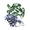
| ||||||||
|---|---|---|---|---|---|---|---|---|---|
| 1 |
| ||||||||
| Unit cell |
|
-Components
| #1: Protein | Mass: 20400.291 Da / Num. of mol.: 2 Source method: isolated from a genetically manipulated source Source: (gene. exp.) References: UniProt: P69503, adenine phosphoribosyltransferase #2: Chemical | ChemComp-MG / |   Mass: 24.305 Da / Num. of mol.: 1 / Source method: obtained synthetically / Formula: Mg Mass: 24.305 Da / Num. of mol.: 1 / Source method: obtained synthetically / Formula: Mg#3: Water | ChemComp-HOH / |  Mass: 18.015 Da / Num. of mol.: 548 / Source method: isolated from a natural source / Formula: H2O Mass: 18.015 Da / Num. of mol.: 548 / Source method: isolated from a natural source / Formula: H2O |
|---|
-Experimental details
-Experiment
| Experiment | Method: X-RAY DIFFRACTION / Number of used crystals: 1 |
|---|
- Sample preparation
| Crystal | Density Matthews: 2.13 Å3/Da / Density % sol: 42.35 % |
|---|---|
| Crystal grow | Temperature: 291 K / Method: microbach / pH: 8.5 Details: 30 w/v(%) PEG 4000, 0.1M Tris, 0.2M Mg Chlor , pH 8.5, Microbach, temperature 291K |
-Data collection
| Diffraction | Mean temperature: 100 K |
|---|---|
| Diffraction source | Source: SYNCHROTRON / Site: SPring-8  / Beamline: BL26B1 / Wavelength: 1 Å / Beamline: BL26B1 / Wavelength: 1 Å |
| Detector | Type: RIGAKU RAXIS V / Detector: IMAGE PLATE / Date: Jul 6, 2006 / Details: mirror |
| Radiation | Monochromator: mirror / Protocol: SINGLE WAVELENGTH / Monochromatic (M) / Laue (L): M / Scattering type: x-ray |
| Radiation wavelength | Wavelength: 1 Å / Relative weight: 1 |
| Reflection | Resolution: 1.25→40 Å / Num. all: 88230 / Num. obs: 88230 / % possible obs: 94.6 % / Observed criterion σ(F): 0 / Observed criterion σ(I): 0 / Redundancy: 3.5 % / Biso Wilson estimate: 11.3 Å2 / Rmerge(I) obs: 0.057 / Rsym value: 0.052 / Net I/σ(I): 9.9 |
| Reflection shell | Resolution: 1.25→1.29 Å / Redundancy: 3.6 % / Rmerge(I) obs: 0.244 / Mean I/σ(I) obs: 3.2 / Rsym value: 0.207 / % possible all: 91.9 |
- Processing
| Software |
| ||||||||||||||||||||||||||||||||||||
|---|---|---|---|---|---|---|---|---|---|---|---|---|---|---|---|---|---|---|---|---|---|---|---|---|---|---|---|---|---|---|---|---|---|---|---|---|---|
| Refinement | Method to determine structure: MOLECULAR REPLACEMENT Starting model: PDB code 1ZN9 Resolution: 1.25→35 Å / Rfactor Rfree error: 0.003 / Data cutoff high absF: 707686.78 / Data cutoff low absF: 0 / Isotropic thermal model: RESTRAINED / Cross valid method: THROUGHOUT / σ(F): 0 / σ(I): 0 / Stereochemistry target values: Engh & Huber
| ||||||||||||||||||||||||||||||||||||
| Solvent computation | Solvent model: FLAT MODEL / Bsol: 34.533 Å2 / ksol: 0.342346 e/Å3 | ||||||||||||||||||||||||||||||||||||
| Displacement parameters | Biso mean: 14.4 Å2
| ||||||||||||||||||||||||||||||||||||
| Refine analyze |
| ||||||||||||||||||||||||||||||||||||
| Refinement step | Cycle: LAST / Resolution: 1.25→35 Å
| ||||||||||||||||||||||||||||||||||||
| Refine LS restraints |
| ||||||||||||||||||||||||||||||||||||
| LS refinement shell | Resolution: 1.25→1.33 Å / Rfactor Rfree error: 0.01 / Total num. of bins used: 6
| ||||||||||||||||||||||||||||||||||||
| Xplor file |
|
