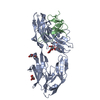
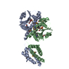
- PDB-5zj6: Crystal structure of HCK kinase complexed with a pyrrolo-pyrimidi... -
+
Open data
ID or keywords:
Loading...
-
Basic information
Entry
Database: PDB / ID: 5zj6
Title
Crystal structure of HCK kinase complexed with a pyrrolo-pyrimidine inhibitor 7-[trans-4-(4-methylpiperazin-1-yl)cyclohexyl]-5-(4-phenoxyphenyl)-7H-pyrrolo[2,3-d]pyrimidin-4-amine
Components
Tyrosine-protein kinase HCK
Keywords
TRANSFERASE / kinase/Kinase inhibitor
Function / homology
Function and homology information
leukocyte degranulation / leukocyte migration involved in immune response / respiratory burst after phagocytosis / regulation of podosome assembly / innate immune response-activating signaling pathway / : / FLT3 signaling through SRC family kinases / regulation of phagocytosis / Nef and signal transduction / Fc-gamma receptor signaling pathway involved in phagocytosis ...leukocyte degranulation / leukocyte migration involved in immune response / respiratory burst after phagocytosis / regulation of podosome assembly / innate immune response-activating signaling pathway / : / FLT3 signaling through SRC family kinases / regulation of phagocytosis / Nef and signal transduction / Fc-gamma receptor signaling pathway involved in phagocytosis / mesoderm development / positive regulation of actin filament polymerization / FCGR activation / type II interferon-mediated signaling pathway / transport vesicle / Signaling by CSF3 (G-CSF) / cell projection / phosphotyrosine residue binding / peptidyl-tyrosine phosphorylation / FCGR3A-mediated IL10 synthesis / lipopolysaccharide-mediated signaling pathway / cell surface receptor protein tyrosine kinase signaling pathway / integrin-mediated signaling pathway / regulation of actin cytoskeleton organization / FCGR3A-mediated phagocytosis / non-specific protein-tyrosine kinase / Regulation of signaling by CBL / negative regulation of inflammatory response to antigenic stimulus / non-membrane spanning protein tyrosine kinase activity / caveola / Inactivation of CSF3 (G-CSF) signaling / cytoplasmic side of plasma membrane / cytokine-mediated signaling pathway / Signaling by CSF1 (M-CSF) in myeloid cells / protein autophosphorylation / regulation of cell shape / protein tyrosine kinase activity / regulation of inflammatory response / cytoskeleton / protein phosphorylation / cell differentiation / lysosome / cell adhesion / intracellular signal transduction / defense response to Gram-positive bacterium / inflammatory response / signaling receptor binding / focal adhesion / positive regulation of cell population proliferation / lipid binding / negative regulation of apoptotic process / Golgi apparatus / ATP binding / nucleus / plasma membrane / cytosol Similarity search - Function
In the structure databanks used in Yorodumi, some data are registered as the other names, "COVID-19 virus" and "2019-nCoV". Here are the details of the virus and the list of structure data.
Jan 31, 2019. EMDB accession codes are about to change! (news from PDBe EMDB page)
EMDB accession codes are about to change! (news from PDBe EMDB page)
The allocation of 4 digits for EMDB accession codes will soon come to an end. Whilst these codes will remain in use, new EMDB accession codes will include an additional digit and will expand incrementally as the available range of codes is exhausted. The current 4-digit format prefixed with “EMD-” (i.e. EMD-XXXX) will advance to a 5-digit format (i.e. EMD-XXXXX), and so on. It is currently estimated that the 4-digit codes will be depleted around Spring 2019, at which point the 5-digit format will come into force.
The EM Navigator/Yorodumi systems omit the EMD- prefix.
Related info.:Q: What is EMD? / ID/Accession-code notation in Yorodumi/EM Navigator
Yorodumi is a browser for structure data from EMDB, PDB, SASBDB, etc.
This page is also the successor to EM Navigator detail page, and also detail information page/front-end page for Omokage search.
The word "yorodu" (or yorozu) is an old Japanese word meaning "ten thousand". "mi" (miru) is to see.
Related info.:EMDB / PDB / SASBDB / Comparison of 3 databanks / Yorodumi Search / Aug 31, 2016. New EM Navigator & Yorodumi / Yorodumi Papers / Jmol/JSmol / Function and homology information / Changes in new EM Navigator and Yorodumi
 Movie
Movie Controller

 Yorodumi
 Open data
 Basic information
 Components
 Keywords
 Function and homology information
 Homo sapiens (human)
 X-RAY DIFFRACTION /
  Authors
 Citation
 Structure visualization
 Downloads & links
 Other downloads
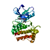


 PDBj
PDBj


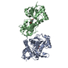
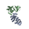
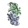
 Assembly
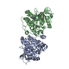

 Spodoptera frugiperda (fall armyworm)
 Mass: 482.620 Da / Num. of mol.: 2 / Source method: obtained synthetically / Formula: C29H34N6O
 Mass: 18.015 Da / Num. of mol.: 676 / Source method: isolated from a natural source / Formula: H2O
 Sample preparation
 / Beamline: BL26B2 / Wavelength: 1 Å
 Processing