 Movie
Movie Controller

[English] 日本語
 Yorodumi
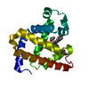
- PDB-5zeo: X-ray structure of sperm whale V21C/V66C/F46S myoglobin mutant wi... -
+ Open data

- Basic information
| Entry | Database: PDB / ID: 5zeo | ||||||
|---|---|---|---|---|---|---|---|
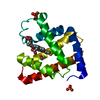
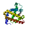
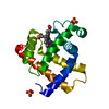
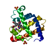
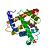
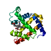
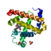
| Title | X-ray structure of sperm whale V21C/V66C/F46S myoglobin mutant with an intramolecular disulfide bond | ||||||
 Components Components | Myoglobin | ||||||
 Keywords Keywords | OXYGEN TRANSPORT / myoglobin / sperm whale | ||||||
| Function / homology |  Function and homology information Function and homology informationnitrite reductase activity / Oxidoreductases; Acting on other nitrogenous compounds as donors / sarcoplasm / Oxidoreductases; Acting on a peroxide as acceptor; Peroxidases / removal of superoxide radicals / oxygen carrier activity / peroxidase activity / oxygen binding / heme binding / extracellular exosome / metal ion binding Similarity search - Function | ||||||
| Biological species |  | ||||||
| Method |  X-RAY DIFFRACTION / SYNCHROTRON / MOLECULAR REPLACEMENT / Resolution: 1.77 Å X-RAY DIFFRACTION / SYNCHROTRON / MOLECULAR REPLACEMENT / Resolution: 1.77 Å | ||||||
 Authors Authors | Yuan, H. | ||||||
 Citation Citation | Journal: Chem. Commun. (Camb.) / Year: 2018 Title: Regulation of both the structure and function by a de novo designed disulfide bond: a case study of heme proteins in myoglobin Authors: Yin, L.L. / Yuan, H. / Du, K.J. / He, B. / Gao, S.Q. / Wen, G.B. / Tan, X. / Lin, Y.W. | ||||||
| History |
|
- Structure visualization
| Structure viewer | Molecule: MolmilJmol/JSmol |
|---|
- Downloads & links
-Download
| PDBx/mmCIF format | 5zeo.cif.gz | 49.7 KB | Display | PDBx/mmCIF format |
|---|---|---|---|---|
| PDB format | pdb5zeo.ent.gz | 33.7 KB | Display | PDB format |
| PDBx/mmJSON format | 5zeo.json.gz | Tree view | PDBx/mmJSON format | |
| Others |  Other downloads Other downloads |
-Validation report
| Arichive directory | https://data.pdbj.org/pub/pdb/validation_reports/ze/5zeoftp://data.pdbj.org/pub/pdb/validation_reports/ze/5zeo | HTTPS FTP |
|---|
-Related structure data
| Related structure data |  5hlxS S: Starting model for refinement |
|---|---|
| Similar structure data |


-Links
 PDBj
PDBj


- Assembly
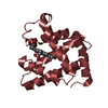
| Deposited unit | 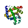
| ||||||||
|---|---|---|---|---|---|---|---|---|---|
| 1 |
| ||||||||
| Unit cell |
|
-Components
| #1: Protein | Mass: 17182.879 Da / Num. of mol.: 1 / Fragment: UNP residues 2-154 / Mutation: V21C, P46S, V66C Source method: isolated from a genetically manipulated source Source: (gene. exp.)  |
|---|---|
| #2: Chemical | ChemComp-HEM /   Mass: 616.487 Da / Num. of mol.: 1 / Source method: obtained synthetically / Formula: C34H32FeN4O4 Mass: 616.487 Da / Num. of mol.: 1 / Source method: obtained synthetically / Formula: C34H32FeN4O4 |
| #3: Water | ChemComp-HOH /  Mass: 18.015 Da / Num. of mol.: 175 / Source method: isolated from a natural source / Formula: H2O Mass: 18.015 Da / Num. of mol.: 175 / Source method: isolated from a natural source / Formula: H2O |
| Has protein modification | Y |
-Experimental details
-Experiment
| Experiment | Method: X-RAY DIFFRACTION / Number of used crystals: 1 |
|---|
- Sample preparation
| Crystal | Density Matthews: 2.16 Å3/Da / Density % sol: 42.96 % |
|---|---|
| Crystal grow | Temperature: 297 K / Method: vapor diffusion, hanging drop Details: 0.2M Sodium acetate trihydrate, 0.1M Sodium cacodylate trihydrate pH 6.5, 30%(w/v) Polyethylene glycol 8,000 |
-Data collection
| Diffraction | Mean temperature: 100 K |
|---|---|
| Diffraction source | Source: SYNCHROTRON / Site: SSRF  / Beamline: BL18U1 / Wavelength: 0.988 Å / Beamline: BL18U1 / Wavelength: 0.988 Å |
| Detector | Type: DECTRIS PILATUS3 S 6M / Detector: PIXEL / Date: Nov 29, 2017 |
| Radiation | Protocol: SINGLE WAVELENGTH / Monochromatic (M) / Laue (L): M / Scattering type: x-ray |
| Radiation wavelength | Wavelength: 0.988 Å / Relative weight: 1 |
| Reflection | Resolution: 1.77→40.63 Å / Num. obs: 15020 / % possible obs: 99.4 % / Redundancy: 5.6 % / Rmerge(I) obs: 0.157 / Net I/σ(I): 23.05 |
| Reflection shell | Resolution: 1.77→1.8 Å / Redundancy: 5.5 % / Rmerge(I) obs: 0.468 / Mean I/σ(I) obs: 5.33 / Num. unique obs: 725 / % possible all: 97.7 |
- Processing
| Software |
| ||||||||||||||||
|---|---|---|---|---|---|---|---|---|---|---|---|---|---|---|---|---|---|
| Refinement | Method to determine structure: MOLECULAR REPLACEMENT Starting model: 5HLX Resolution: 1.77→40.63 Å / Cross valid method: FREE R-VALUE
| ||||||||||||||||
| Refinement step | Cycle: LAST / Resolution: 1.77→40.63 Å
|
