 Movie
Movie Controller

[English] 日本語
 Yorodumi
- PDB-5zcy: Crystal structure of archaeal translation initiation factor 1 at ... -
+ Open data

- Basic information
| Entry | Database: PDB / ID: 5zcy | ||||||
|---|---|---|---|---|---|---|---|
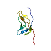
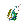
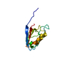
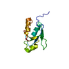
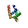
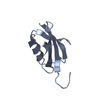
| Title | Crystal structure of archaeal translation initiation factor 1 at 1.5 Angstroms resolution | ||||||
 Components Components | Protein translation factor SUI1 homolog | ||||||
 Keywords Keywords | TRANSLATION / aIF1 / eIF1 / eukaryotic translation initiation / YciH / archaea / eukaryote | ||||||
| Function / homology |  Function and homology information Function and homology information | ||||||
| Biological species |   Pyrococcus horikoshii (archaea) Pyrococcus horikoshii (archaea) | ||||||
| Method |  X-RAY DIFFRACTION / MOLECULAR REPLACEMENT / molecular replacement / Resolution: 1.5 Å X-RAY DIFFRACTION / MOLECULAR REPLACEMENT / molecular replacement / Resolution: 1.5 Å | ||||||
 Authors Authors | Gogoi, P. / Kanaujia, S.P. | ||||||
| Funding support |  India, 1items India, 1items
| ||||||
 Citation Citation | Journal: FEBS Lett. / Year: 2018 Title: Archaeal and eukaryal translation initiation factor 1 differ in their RNA interacting loops. Authors: Gogoi, P. / Kanaujia, S.P. | ||||||
| History |
|
- Structure visualization
| Structure viewer | Molecule: MolmilJmol/JSmol |
|---|
- Downloads & links
-Download
| PDBx/mmCIF format | 5zcy.cif.gz | 33.3 KB | Display | PDBx/mmCIF format |
|---|---|---|---|---|
| PDB format | pdb5zcy.ent.gz | 20.7 KB | Display | PDB format |
| PDBx/mmJSON format | 5zcy.json.gz | Tree view | PDBx/mmJSON format | |
| Others |  Other downloads Other downloads |
-Validation report
| Arichive directory | https://data.pdbj.org/pub/pdb/validation_reports/zc/5zcyftp://data.pdbj.org/pub/pdb/validation_reports/zc/5zcy | HTTPS FTP |
|---|
-Related structure data
| Related structure data | 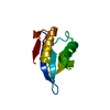 4mo0S S: Starting model for refinement |
|---|---|
| Similar structure data |


-Links
 PDBj
PDBj- Assembly
| Deposited unit | 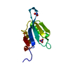
| ||||||||
|---|---|---|---|---|---|---|---|---|---|
| 1 |
| ||||||||
| Unit cell |
|
-Components
| #1: Protein | Mass: 11474.438 Da / Num. of mol.: 1 Source method: isolated from a genetically manipulated source Source: (gene. exp.) Pyrococcus horikoshii (strain ATCC 700860 / DSM 12428 / JCM 9974 / NBRC 100139 / OT-3) (archaea)Strain: ATCC 700860 / DSM 12428 / JCM 9974 / NBRC 100139 / OT-3 Gene: PH1771.1 / Plasmid: pET22b / Production host:  |
|---|---|
| #2: Chemical | ChemComp-NO3 /   Mass: 62.005 Da / Num. of mol.: 1 / Source method: obtained synthetically / Formula: NO3 Mass: 62.005 Da / Num. of mol.: 1 / Source method: obtained synthetically / Formula: NO3 |
| #3: Chemical | ChemComp-NO2 /   Mass: 46.005 Da / Num. of mol.: 1 / Source method: obtained synthetically / Formula: NO2 Mass: 46.005 Da / Num. of mol.: 1 / Source method: obtained synthetically / Formula: NO2 |
| #4: Water | ChemComp-HOH /  Mass: 18.015 Da / Num. of mol.: 82 / Source method: isolated from a natural source / Formula: H2O Mass: 18.015 Da / Num. of mol.: 82 / Source method: isolated from a natural source / Formula: H2O |
-Experimental details
-Experiment
| Experiment | Method: X-RAY DIFFRACTION / Number of used crystals: 1 |
|---|
- Sample preparation
| Crystal | Density Matthews: 2.06 Å3/Da / Density % sol: 40.18 % |
|---|---|
| Crystal grow | Temperature: 293 K / Method: microbatch / Details: 0.2M Lithium Nitrate, 20% PEG 3350 |
-Data collection
| Diffraction | Mean temperature: 100 K |
|---|---|
| Diffraction source | Source: ROTATING ANODE / Type: RIGAKU MICROMAX-007 HF / Wavelength: 1.5418 Å |
| Detector | Type: RIGAKU RAXIS IV++ / Detector: IMAGE PLATE / Date: Oct 26, 2017 / Details: VariMax HF |
| Radiation | Protocol: SINGLE WAVELENGTH / Monochromatic (M) / Laue (L): M / Scattering type: x-ray |
| Radiation wavelength | Wavelength: 1.5418 Å / Relative weight: 1 |
| Reflection | Resolution: 1.5→35.97 Å / Num. obs: 14801 / % possible obs: 98.1 % / Redundancy: 11 % / CC1/2: 1 / Rmerge(I) obs: 0.054 / Rpim(I) all: 0.017 / Rrim(I) all: 0.056 / Net I/σ(I): 23.4 |
| Reflection shell | Resolution: 1.5→1.52 Å / Redundancy: 10.3 % / Rmerge(I) obs: 0.514 / Num. unique obs: 691 / CC1/2: 0.915 / Rpim(I) all: 0.165 / Rrim(I) all: 0.54 / % possible all: 95.9 |
-Phasing
| Phasing | Method: molecular replacement | |||||||||
|---|---|---|---|---|---|---|---|---|---|---|
| Phasing MR | Model details: Phaser MODE: MR_AUTO
|
- Processing
| Software |
| ||||||||||||||||||||||||||||||||||||||||||||||||||||||||||||
|---|---|---|---|---|---|---|---|---|---|---|---|---|---|---|---|---|---|---|---|---|---|---|---|---|---|---|---|---|---|---|---|---|---|---|---|---|---|---|---|---|---|---|---|---|---|---|---|---|---|---|---|---|---|---|---|---|---|---|---|---|---|
| Refinement | Method to determine structure: MOLECULAR REPLACEMENT Starting model: 4MO0 Resolution: 1.5→35.97 Å / Cor.coef. Fo:Fc: 0.962 / Cor.coef. Fo:Fc free: 0.949 / SU B: 1.083 / SU ML: 0.042 / SU R Cruickshank DPI: 0.0696 / Cross valid method: THROUGHOUT / σ(F): 0 / ESU R: 0.07 / ESU R Free: 0.073 Details: HYDROGENS HAVE BEEN ADDED IN THE RIDING POSITIONS U VALUES : REFINED INDIVIDUALLY
| ||||||||||||||||||||||||||||||||||||||||||||||||||||||||||||
| Solvent computation | Ion probe radii: 0.8 Å / Shrinkage radii: 0.8 Å / VDW probe radii: 1.2 Å | ||||||||||||||||||||||||||||||||||||||||||||||||||||||||||||
| Displacement parameters | Biso max: 64.26 Å2 / Biso mean: 19.198 Å2 / Biso min: 7.7 Å2
| ||||||||||||||||||||||||||||||||||||||||||||||||||||||||||||
| Refinement step | Cycle: final / Resolution: 1.5→35.97 Å
| ||||||||||||||||||||||||||||||||||||||||||||||||||||||||||||
| Refine LS restraints |
| ||||||||||||||||||||||||||||||||||||||||||||||||||||||||||||
| LS refinement shell | Resolution: 1.495→1.534 Å / Rfactor Rfree error: 0 / Total num. of bins used: 20
|
