 Movie
Movie Controller

[English] 日本語
 Yorodumi
- PDB-1ihj: Crystal Structure of the N-terminal PDZ domain of InaD in complex... -
+ Open data

- Basic information
| Entry | Database: PDB / ID: 1ihj | ||||||
|---|---|---|---|---|---|---|---|
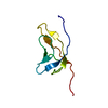
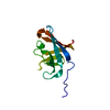
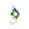
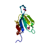
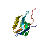
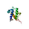
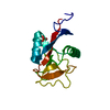
| Title | Crystal Structure of the N-terminal PDZ domain of InaD in complex with a NorpA C-terminal peptide | ||||||
 Components Components |
| ||||||
 Keywords Keywords | SIGNALING PROTEIN / intermolecular disulfide bond / PDZ domain | ||||||
| Function / homology |  Function and homology information Function and homology informationmyosin III binding / detection of light stimulus involved in sensory perception / inaD signaling complex / negative regulation of opsin-mediated signaling pathway / Antigen processing: Ubiquitination & Proteasome degradation / rhabdomere / cellular response to light stimulus / myosin binding / photoreceptor activity / phototransduction ...myosin III binding / detection of light stimulus involved in sensory perception / inaD signaling complex / negative regulation of opsin-mediated signaling pathway / Antigen processing: Ubiquitination & Proteasome degradation / rhabdomere / cellular response to light stimulus / myosin binding / photoreceptor activity / phototransduction / visual perception / sensory perception of sound / intracellular protein localization / signaling receptor complex adaptor activity / calmodulin binding Similarity search - Function | ||||||
| Biological species |  | ||||||
| Method |  X-RAY DIFFRACTION / SYNCHROTRON / MOLECULAR REPLACEMENT / Resolution: 1.8 Å X-RAY DIFFRACTION / SYNCHROTRON / MOLECULAR REPLACEMENT / Resolution: 1.8 Å | ||||||
 Authors Authors | Kimple, M.E. / Siderovski, D.P. / Sondek, J. | ||||||
 Citation Citation | Journal: EMBO J. / Year: 2001 Title: Functional relevance of the disulfide-linked complex of the N-terminal PDZ domain of InaD with NorpA. Authors: Kimple, M.E. / Siderovski, D.P. / Sondek, J. | ||||||
| History |
|
- Structure visualization
| Structure viewer | Molecule: MolmilJmol/JSmol |
|---|
- Downloads & links
-Download
| PDBx/mmCIF format | 1ihj.cif.gz | 53.5 KB | Display | PDBx/mmCIF format |
|---|---|---|---|---|
| PDB format | pdb1ihj.ent.gz | 38.7 KB | Display | PDB format |
| PDBx/mmJSON format | 1ihj.json.gz | Tree view | PDBx/mmJSON format | |
| Others |  Other downloads Other downloads |
-Validation report
| Arichive directory | https://data.pdbj.org/pub/pdb/validation_reports/ih/1ihjftp://data.pdbj.org/pub/pdb/validation_reports/ih/1ihj | HTTPS FTP |
|---|
-Related structure data
| Related structure data |  1be9S S: Starting model for refinement |
|---|---|
| Similar structure data |
-Links
 PDBj
PDBj- Assembly
| Deposited unit | 
| ||||||||
|---|---|---|---|---|---|---|---|---|---|
| 1 | 
| ||||||||
| 2 | 
| ||||||||
| Unit cell |
|
-Components
| #1: Protein | Mass: 10771.533 Da / Num. of mol.: 2 / Fragment: PDZ1 domain Source method: isolated from a genetically manipulated source Source: (gene. exp.)  #2: Protein/peptide | Mass: 755.859 Da / Num. of mol.: 2 / Fragment: C-terminus / Source method: obtained synthetically Details: This peptide was chemically synthesized. It consists of the final seven residues of phospholipase C (gktefca). References: GenBank: 85099, phospholipase C #3: Water | ChemComp-HOH / |  Mass: 18.015 Da / Num. of mol.: 172 / Source method: isolated from a natural source / Formula: H2O Mass: 18.015 Da / Num. of mol.: 172 / Source method: isolated from a natural source / Formula: H2OHas protein modification | Y | |
|---|
-Experimental details
-Experiment
| Experiment | Method: X-RAY DIFFRACTION / Number of used crystals: 1 |
|---|
- Sample preparation
| Crystal | Density Matthews: 2.81 Å3/Da / Density % sol: 56.17 % | ||||||||||||||||||||||||||||||||||||
|---|---|---|---|---|---|---|---|---|---|---|---|---|---|---|---|---|---|---|---|---|---|---|---|---|---|---|---|---|---|---|---|---|---|---|---|---|---|
| Crystal grow | Temperature: 300 K / Method: vapor diffusion, sitting drop / pH: 8.5 Details: PEG 4000, lithium sulfate, glycerol, dithiothreitol, pH 8.5, VAPOR DIFFUSION, SITTING DROP, temperature 300K | ||||||||||||||||||||||||||||||||||||
| Crystal grow | *PLUS Details: used seeding | ||||||||||||||||||||||||||||||||||||
| Components of the solutions | *PLUS
|
-Data collection
| Diffraction | Mean temperature: 100 K |
|---|---|
| Diffraction source | Source: SYNCHROTRON / Site: NSLS  / Beamline: X4A / Wavelength: 1.0072 Å / Beamline: X4A / Wavelength: 1.0072 Å |
| Detector | Type: ADSC QUANTUM 4 / Detector: CCD / Date: Aug 19, 2000 / Details: mirrors |
| Radiation | Protocol: SINGLE WAVELENGTH / Monochromatic (M) / Laue (L): M / Scattering type: x-ray |
| Radiation wavelength | Wavelength: 1.0072 Å / Relative weight: 1 |
| Reflection | Resolution: 1.76→20 Å / Num. all: 27241 / Num. obs: 25906 / % possible obs: 95.1 % / Observed criterion σ(F): 1.5 / Observed criterion σ(I): 1.5 / Redundancy: 3.6 % / Biso Wilson estimate: 14.7 Å2 / Rmerge(I) obs: 0.059 / Net I/σ(I): 17.5 |
| Reflection shell | Resolution: 1.76→1.83 Å / Redundancy: 2.9 % / Rmerge(I) obs: 0.156 / % possible all: 86.7 |
| Reflection | *PLUS Lowest resolution: 20 Å |
| Reflection shell | *PLUS % possible obs: 86.7 % / Num. unique obs: 2424 / Mean I/σ(I) obs: 8.5 |
- Processing
| Software |
| ||||||||||||||||||||||||||||||
|---|---|---|---|---|---|---|---|---|---|---|---|---|---|---|---|---|---|---|---|---|---|---|---|---|---|---|---|---|---|---|---|
| Refinement | Method to determine structure: MOLECULAR REPLACEMENT Starting model: PDB Entry 1BE9 minus peptide Resolution: 1.8→20 Å / Cross valid method: THROUGHOUT / σ(F): 2 / σ(I): 2 / Stereochemistry target values: Engh & Huber
| ||||||||||||||||||||||||||||||
| Refinement step | Cycle: LAST / Resolution: 1.8→20 Å
| ||||||||||||||||||||||||||||||
| Software | *PLUS Name: CNS / Classification: refinement | ||||||||||||||||||||||||||||||
| Refinement | *PLUS Highest resolution: 1.8 Å / Lowest resolution: 20 Å / σ(F): 2 | ||||||||||||||||||||||||||||||
| Solvent computation | *PLUS | ||||||||||||||||||||||||||||||
| Displacement parameters | *PLUS | ||||||||||||||||||||||||||||||
| Refine LS restraints | *PLUS
|
