 Movie
Movie Controller


+ Open data

- Basic information
| Entry | Database: PDB / ID: 5zbj | ||||||
|---|---|---|---|---|---|---|---|
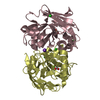
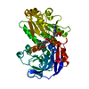
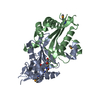
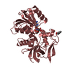
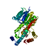
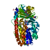
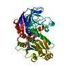
| Title | Crystal structure of type-I LOG from Pseudomonas aeruginosa PAO1 | ||||||
 Components Components | Putative cytokinin riboside 5'-monophosphate phosphoribohydrolase | ||||||
 Keywords Keywords | HYDROLASE | ||||||
| Function / homology |  Function and homology information Function and homology informationcytokinin riboside 5'-monophosphate phosphoribohydrolase activity / : / AMP nucleosidase / AMP nucleosidase activity / hydrolase activity, hydrolyzing N-glycosyl compounds / cytokinin biosynthetic process / cytosol Similarity search - Function | ||||||
| Biological species |   Pseudomonas aeruginosa (bacteria) Pseudomonas aeruginosa (bacteria) | ||||||
| Method |  X-RAY DIFFRACTION / SYNCHROTRON / MOLECULAR REPLACEMENT / Resolution: 1.89 Å X-RAY DIFFRACTION / SYNCHROTRON / MOLECULAR REPLACEMENT / Resolution: 1.89 Å | ||||||
 Authors Authors | Seo, H. / Kim, K.-J. | ||||||
 Citation Citation | Journal: Environ. Microbiol. / Year: 2018 Title: Structural insight into molecular mechanism of cytokinin activating protein from Pseudomonas aeruginosa PAO1. Authors: Seo, H. / Kim, K.J. | ||||||
| History |
|
- Structure visualization
| Structure viewer | Molecule: MolmilJmol/JSmol |
|---|
- Downloads & links
-Download
| PDBx/mmCIF format | 5zbj.cif.gz | 53.8 KB | Display | PDBx/mmCIF format |
|---|---|---|---|---|
| PDB format | pdb5zbj.ent.gz | 36.5 KB | Display | PDB format |
| PDBx/mmJSON format | 5zbj.json.gz | Tree view | PDBx/mmJSON format | |
| Others |  Other downloads Other downloads |
-Validation report
| Summary document | 5zbj_validation.pdf.gz | 468.8 KB | Display | wwPDB validaton report |
|---|---|---|---|---|
| Full document | 5zbj_full_validation.pdf.gz | 468.8 KB | Display | |
| Data in XML | 5zbj_validation.xml.gz | 10.3 KB | Display | |
| Data in CIF | 5zbj_validation.cif.gz | 14 KB | Display | |
| Arichive directory | https://data.pdbj.org/pub/pdb/validation_reports/zb/5zbjftp://data.pdbj.org/pub/pdb/validation_reports/zb/5zbj | HTTPS FTP |
-Related structure data
| Related structure data |  5zbkC  5zblC  5itsS S: Starting model for refinement C: citing same article ( |
|---|---|
| Similar structure data |


-Links
 PDBj
PDBj


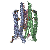
- Assembly
| Deposited unit | 
| ||||||||
|---|---|---|---|---|---|---|---|---|---|
| 1 | 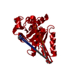
| ||||||||
| Unit cell |
|
-Components
-Protein , 1 types, 1 molecules A
| #1: Protein | Mass: 21816.969 Da / Num. of mol.: 1 Source method: isolated from a genetically manipulated source Source: (gene. exp.) Pseudomonas aeruginosa (strain ATCC 15692 / DSM 22644 / CIP 104116 / JCM 14847 / LMG 12228 / 1C / PRS 101 / PAO1) (bacteria)Strain: ATCC 15692 / DSM 22644 / CIP 104116 / JCM 14847 / LMG 12228 / 1C / PRS 101 / PAO1 Gene: PA4923 / Plasmid: pET30a / Production host: References: UniProt: P48636, Hydrolases; Glycosylases; Hydrolysing N-glycosyl compounds |
|---|
-Non-polymers , 5 types, 106 molecules 


| #2: Chemical |  Mass: 92.094 Da / Num. of mol.: 3 / Source method: obtained synthetically / Formula: C3H8O3 Mass: 92.094 Da / Num. of mol.: 3 / Source method: obtained synthetically / Formula: C3H8O3#3: Chemical | ChemComp-PO4 / |  Mass: 94.971 Da / Num. of mol.: 1 / Source method: obtained synthetically / Formula: PO4 Mass: 94.971 Da / Num. of mol.: 1 / Source method: obtained synthetically / Formula: PO4#4: Chemical | ChemComp-IMD / |  Mass: 69.085 Da / Num. of mol.: 1 / Source method: obtained synthetically / Formula: C3H5N2 Mass: 69.085 Da / Num. of mol.: 1 / Source method: obtained synthetically / Formula: C3H5N2#5: Chemical | ChemComp-EDO / |  Mass: 62.068 Da / Num. of mol.: 1 / Source method: obtained synthetically / Formula: C2H6O2 Mass: 62.068 Da / Num. of mol.: 1 / Source method: obtained synthetically / Formula: C2H6O2#6: Water | ChemComp-HOH / | Mass: 18.015 Da / Num. of mol.: 100 / Source method: isolated from a natural source / Formula: H2O |
|---|
-Experimental details
-Experiment
| Experiment | Method: X-RAY DIFFRACTION / Number of used crystals: 1 |
|---|
- Sample preparation
| Crystal | Density Matthews: 2.61 Å3/Da / Density % sol: 52.88 % |
|---|---|
| Crystal grow | Temperature: 293 K / Method: vapor diffusion, hanging drop / pH: 6.8 / Details: ammonium phosphate dibasic, imidazole pH 8.0 |
-Data collection
| Diffraction | Mean temperature: 100 K |
|---|---|
| Diffraction source | Source: SYNCHROTRON / Site: PAL/PLS  / Beamline: 7A (6B, 6C1) / Wavelength: 0.97934 Å / Beamline: 7A (6B, 6C1) / Wavelength: 0.97934 Å |
| Detector | Type: ADSC QUANTUM 270 / Detector: CCD / Date: Feb 28, 2017 |
| Radiation | Monochromator: Double Crystal Monochromator / Protocol: SINGLE WAVELENGTH / Monochromatic (M) / Laue (L): M / Scattering type: x-ray |
| Radiation wavelength | Wavelength: 0.97934 Å / Relative weight: 1 |
| Reflection | Resolution: 1.89→54.11 Å / Num. obs: 18128 / % possible obs: 98.1 % / Redundancy: 5 % / CC1/2: 0.804 / Rmerge(I) obs: 0.072 / Rpim(I) all: 0.036 / Net I/σ(I): 4.5 |
| Reflection shell | Resolution: 1.89→1.93 Å / Rmerge(I) obs: 0.347 / Num. unique obs: 845 / CC1/2: 0.944 / Rpim(I) all: 0.158 |
- Processing
| Software |
| ||||||||||||||||||||||||||||||||||||||||||||||||||||||||||||
|---|---|---|---|---|---|---|---|---|---|---|---|---|---|---|---|---|---|---|---|---|---|---|---|---|---|---|---|---|---|---|---|---|---|---|---|---|---|---|---|---|---|---|---|---|---|---|---|---|---|---|---|---|---|---|---|---|---|---|---|---|---|
| Refinement | Method to determine structure: MOLECULAR REPLACEMENT Starting model: 5ITS Resolution: 1.89→54.11 Å / Cor.coef. Fo:Fc: 0.966 / Cor.coef. Fo:Fc free: 0.94 / SU B: 2.718 / SU ML: 0.078 / Cross valid method: THROUGHOUT / σ(F): 0 / ESU R: 0.109 / ESU R Free: 0.118 Details: HYDROGENS HAVE BEEN ADDED IN THE RIDING POSITIONS U VALUES : REFINED INDIVIDUALLY
| ||||||||||||||||||||||||||||||||||||||||||||||||||||||||||||
| Solvent computation | Ion probe radii: 0.8 Å / Shrinkage radii: 0.8 Å / VDW probe radii: 1.2 Å | ||||||||||||||||||||||||||||||||||||||||||||||||||||||||||||
| Displacement parameters | Biso max: 76.68 Å2 / Biso mean: 22.512 Å2 / Biso min: 10.87 Å2
| ||||||||||||||||||||||||||||||||||||||||||||||||||||||||||||
| Refinement step | Cycle: final / Resolution: 1.89→54.11 Å
| ||||||||||||||||||||||||||||||||||||||||||||||||||||||||||||
| Refine LS restraints |
| ||||||||||||||||||||||||||||||||||||||||||||||||||||||||||||
| LS refinement shell | Resolution: 1.894→1.943 Å / Rfactor Rfree error: 0 / Total num. of bins used: 20
|
