 Movie
Movie Controller

[English] 日本語
 Yorodumi
- PDB-5z74: Crystal structure of alkaline/neutral invertase InvB from Anabaen... -
+ Open data

- Basic information
| Entry | Database: PDB / ID: 5z74 | |||||||||
|---|---|---|---|---|---|---|---|---|---|---|
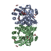
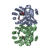
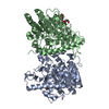
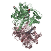
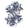
| Title | Crystal structure of alkaline/neutral invertase InvB from Anabaena sp. PCC 7120 complexed with sucrose | |||||||||
 Components Components | Alr0819 protein | |||||||||
 Keywords Keywords | HYDROLASE / alkaline/neutral invertases / cyanobacteria / glycoside hydrolase family 100 / sucrose hydrolysis | |||||||||
| Function / homology |  Function and homology information Function and homology informationendo-alpha-N-acetylgalactosaminidase activity / beta-fructofuranosidase / sucrose catabolic process / sucrose alpha-glucosidase activity Similarity search - Function | |||||||||
| Biological species |  Nostoc sp. (bacteria) Nostoc sp. (bacteria) | |||||||||
| Method |  X-RAY DIFFRACTION / SYNCHROTRON / MOLECULAR REPLACEMENT / Resolution: 1.95 Å X-RAY DIFFRACTION / SYNCHROTRON / MOLECULAR REPLACEMENT / Resolution: 1.95 Å | |||||||||
 Authors Authors | Xie, J. / Hu, H.X. / Cai, K. / Yang, F. / Jiang, Y.L. / Chen, Y. / Zhou, C.Z. | |||||||||
| Funding support |  China, 1items China, 1items
| |||||||||
 Citation Citation | Journal: FEBS Lett. / Year: 2018 Title: Structural and enzymatic analyses of Anabaena heterocyst-specific alkaline invertase InvB. Authors: Xie, J. / Hu, H.X. / Cai, K. / Xia, L.Y. / Yang, F. / Jiang, Y.L. / Chen, Y. / Zhou, C.Z. | |||||||||
| History |
|
- Structure visualization
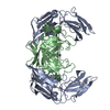
| Structure viewer | Molecule: MolmilJmol/JSmol |
|---|
- Downloads & links
-Download
| PDBx/mmCIF format | 5z74.cif.gz | 189.4 KB | Display | PDBx/mmCIF format |
|---|---|---|---|---|
| PDB format | pdb5z74.ent.gz | 150.2 KB | Display | PDB format |
| PDBx/mmJSON format | 5z74.json.gz | Tree view | PDBx/mmJSON format | |
| Others |  Other downloads Other downloads |
-Validation report
| Arichive directory | https://data.pdbj.org/pub/pdb/validation_reports/z7/5z74ftp://data.pdbj.org/pub/pdb/validation_reports/z7/5z74 | HTTPS FTP |
|---|
-Related structure data
| Related structure data |  5z73SC S: Starting model for refinement C: citing same article ( |
|---|---|
| Similar structure data |


-Links
 PDBj
PDBj
- Assembly
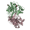
| Deposited unit | 
| ||||||||
|---|---|---|---|---|---|---|---|---|---|
| 1 |
| ||||||||
| Unit cell |
|
-Components
| #1: Protein | Mass: 52609.516 Da / Num. of mol.: 2 Source method: isolated from a genetically manipulated source Source: (gene. exp.) Nostoc sp. (strain PCC 7120 / SAG 25.82 / UTEX 2576) (bacteria)Strain: PCC 7120 / SAG 25.82 / UTEX 2576 / Gene: invB, alr0819 / Production host: #2: Polysaccharide |   Source method: isolated from a genetically manipulated source Details: oligosaccharide with reducing-end-to-reducing-end glycosidic bond References: sucrose #3: Water | ChemComp-HOH / |  Mass: 18.015 Da / Num. of mol.: 184 / Source method: isolated from a natural source / Formula: H2O Mass: 18.015 Da / Num. of mol.: 184 / Source method: isolated from a natural source / Formula: H2OHas protein modification | Y | |
|---|
-Experimental details
-Experiment
| Experiment | Method: X-RAY DIFFRACTION / Number of used crystals: 1 |
|---|
- Sample preparation
| Crystal | Density Matthews: 2.77 Å3/Da / Density % sol: 55.67 % |
|---|---|
| Crystal grow | Temperature: 289 K / Method: vapor diffusion, hanging drop / pH: 6 Details: 0.7M lithium chloride, 6% polyethylene glycol 6000, 0.1M MES, pH 6.0 |
-Data collection
| Diffraction | Mean temperature: 100 K |
|---|---|
| Diffraction source | Source: SYNCHROTRON / Site: SSRF / Beamline: BL17U1 / Wavelength: 0.97916 Å |
| Detector | Type: ADSC QUANTUM 315r / Detector: CCD / Date: Mar 27, 2016 |
| Radiation | Monochromator: SAGITALLY FOCUSED Si(111) / Protocol: SINGLE WAVELENGTH / Monochromatic (M) / Laue (L): M / Scattering type: x-ray |
| Radiation wavelength | Wavelength: 0.97916 Å / Relative weight: 1 |
| Reflection | Resolution: 1.95→50 Å / Num. obs: 80206 / % possible obs: 95.7 % / Redundancy: 3.5 % / Rmerge(I) obs: 0.096 / Net I/σ(I): 13.1 |
| Reflection shell | Resolution: 1.95→2.02 Å / Redundancy: 3.5 % / Rmerge(I) obs: 0.537 / Mean I/σ(I) obs: 2.4 / Num. unique obs: 8173 / % possible all: 98.1 |
- Processing
| Software |
| ||||||||||||||||||||||||||||||||||||||||||||||||||||||||||||||||||||||||||||||||||||||||||||||||||||||||||||||||||||||||||||||||||||||||||||||||||||||||||||||||||||||||||||||||||||||
|---|---|---|---|---|---|---|---|---|---|---|---|---|---|---|---|---|---|---|---|---|---|---|---|---|---|---|---|---|---|---|---|---|---|---|---|---|---|---|---|---|---|---|---|---|---|---|---|---|---|---|---|---|---|---|---|---|---|---|---|---|---|---|---|---|---|---|---|---|---|---|---|---|---|---|---|---|---|---|---|---|---|---|---|---|---|---|---|---|---|---|---|---|---|---|---|---|---|---|---|---|---|---|---|---|---|---|---|---|---|---|---|---|---|---|---|---|---|---|---|---|---|---|---|---|---|---|---|---|---|---|---|---|---|---|---|---|---|---|---|---|---|---|---|---|---|---|---|---|---|---|---|---|---|---|---|---|---|---|---|---|---|---|---|---|---|---|---|---|---|---|---|---|---|---|---|---|---|---|---|---|---|---|---|
| Refinement | Method to determine structure: MOLECULAR REPLACEMENT Starting model: 5Z73 Resolution: 1.95→31.35 Å / Cor.coef. Fo:Fc: 0.954 / Cor.coef. Fo:Fc free: 0.938 / SU B: 3.686 / SU ML: 0.104 / Cross valid method: THROUGHOUT / ESU R: 0.165 / ESU R Free: 0.148 / Details: HYDROGENS HAVE BEEN ADDED IN THE RIDING POSITIONS
| ||||||||||||||||||||||||||||||||||||||||||||||||||||||||||||||||||||||||||||||||||||||||||||||||||||||||||||||||||||||||||||||||||||||||||||||||||||||||||||||||||||||||||||||||||||||
| Solvent computation | Ion probe radii: 0.8 Å / Shrinkage radii: 0.8 Å / VDW probe radii: 1.2 Å | ||||||||||||||||||||||||||||||||||||||||||||||||||||||||||||||||||||||||||||||||||||||||||||||||||||||||||||||||||||||||||||||||||||||||||||||||||||||||||||||||||||||||||||||||||||||
| Displacement parameters | Biso mean: 37.03 Å2
| ||||||||||||||||||||||||||||||||||||||||||||||||||||||||||||||||||||||||||||||||||||||||||||||||||||||||||||||||||||||||||||||||||||||||||||||||||||||||||||||||||||||||||||||||||||||
| Refinement step | Cycle: 1 / Resolution: 1.95→31.35 Å
| ||||||||||||||||||||||||||||||||||||||||||||||||||||||||||||||||||||||||||||||||||||||||||||||||||||||||||||||||||||||||||||||||||||||||||||||||||||||||||||||||||||||||||||||||||||||
| Refine LS restraints |
|