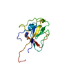
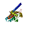
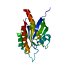
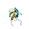
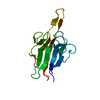
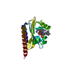
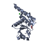
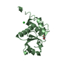
[English] 日本語
Yorodumi
- PDB-5z5b: Crystal structure of Tk-PTP in the G95A mutant form -

+
Open data


ID or keywords:

Loading...

-
Basic information

Entry
Database: PDB / ID: 5z5b
TitleCrystal structure of Tk-PTP in the G95A mutant form
ComponentsProtein-tyrosine phosphatase
KeywordsHYDROLASE / tyrosine phosphatase
Function / homology
Function and homology information


protein tyrosine/serine/threonine phosphatase activity / dephosphorylation / protein-tyrosine-phosphatase / protein tyrosine phosphatase activity / cytoplasm
Similarity search - Function
Dual specificity phosphatase, catalytic domain / Dual specificity phosphatase, catalytic domain / Dual specificity phosphatase, catalytic domain / Dual specificity protein phosphatase domain / Dual specificity protein phosphatase domain profile. / Protein tyrosine phosphatase superfamily / Protein-Tyrosine Phosphatase; Chain A / PTP type protein phosphatase domain profile. / Tyrosine-specific protein phosphatase, PTPase domain / Protein-tyrosine phosphatase, catalytic ...Dual specificity phosphatase, catalytic domain / Dual specificity phosphatase, catalytic domain / Dual specificity phosphatase, catalytic domain / Dual specificity protein phosphatase domain / Dual specificity protein phosphatase domain profile. / Protein tyrosine phosphatase superfamily / Protein-Tyrosine Phosphatase; Chain A / PTP type protein phosphatase domain profile. / Tyrosine-specific protein phosphatase, PTPase domain / Protein-tyrosine phosphatase, catalytic / Protein tyrosine phosphatase, catalytic domain motif / Tyrosine specific protein phosphatases active site. / Protein-tyrosine phosphatase, active site / Tyrosine-specific protein phosphatases domain / Tyrosine specific protein phosphatases domain profile. / Protein-tyrosine phosphatase-like / Alpha-Beta Complex / Alpha Beta
Similarity search - Domain/homology
FORMIC ACID / protein-tyrosine-phosphatase
Similarity search - Component
Biological speciesThermococcus kodakarensis KOD1 (archaea)
MethodX-RAY DIFFRACTION / SYNCHROTRON / MOLECULAR REPLACEMENT / Resolution: 2.3 Å
AuthorsKu, B. / Yun, H.Y. / Kim, S.J.
CitationJournal: PLoS ONE / Year: 2018
Title: Structural study reveals the temperature-dependent conformational flexibility of Tk-PTP, a protein tyrosine phosphatase from Thermococcus kodakaraensis KOD1
Authors: Yun, H.Y. / Lee, J. / Kim, H. / Ryu, H. / Shin, H.C. / Oh, B.H. / Ku, B. / Kim, S.J.
History
DepositionJan 17, 2018Deposition site: PDBJ / Processing site: PDBJ
Revision 1.0Jun 27, 2018Provider: repository / Type: Initial release
Revision 1.1Nov 22, 2023Group: Data collection / Database references / Refinement description
Category: chem_comp_atom / chem_comp_bond ...chem_comp_atom / chem_comp_bond / database_2 / pdbx_initial_refinement_model
Item: _database_2.pdbx_DOI / _database_2.pdbx_database_accession

-
Structure visualization

Structure viewerMolecule:
MolmilJmol/JSmol

Downloads & links

-
Assembly

Deposited unit
A: Protein-tyrosine phosphatase
B: Protein-tyrosine phosphatase
C: Protein-tyrosine phosphatase
hetero molecules


Theoretical massNumber of molelcules
Total (without water)55,7648
Polymers55,5553
Non-polymers2095
Water0
1
A: Protein-tyrosine phosphatase
hetero molecules


Theoretical massNumber of molelcules
Total (without water)18,6003
Polymers18,5181
Non-polymers812
Water0
TypeNameSymmetry operationNumber
identity operation1_555x,y,z1
Buried area80 Å2
ΔGint-5 kcal/mol
Surface area7720 Å2
MethodPISA
2
B: Protein-tyrosine phosphatase
hetero molecules


Theoretical massNumber of molelcules
Total (without water)18,6003
Polymers18,5181
Non-polymers812
Water0
TypeNameSymmetry operationNumber
identity operation1_555x,y,z1
Buried area110 Å2
ΔGint-7 kcal/mol
Surface area7850 Å2
MethodPISA
3
C: Protein-tyrosine phosphatase
hetero molecules


Theoretical massNumber of molelcules
Total (without water)18,5642
Polymers18,5181
Non-polymers461
Water0
TypeNameSymmetry operationNumber
identity operation1_555x,y,z1
Buried area0 Å2
ΔGint0 kcal/mol
Surface area7290 Å2
MethodPISA
Unit cell
Length a, b, c (Å)154.796, 154.796, 75.240
Angle α, β, γ (deg.)90.00, 90.00, 120.00
Int Tables number146
Space group name H-MH3

-
Components

#1: Protein Protein-tyrosine phosphatase


Mass: 18518.312 Da / Num. of mol.: 3 / Mutation: G95A
Source method: isolated from a genetically manipulated source
Source: (gene. exp.) Thermococcus kodakarensis KOD1 (archaea)
Strain: KOD1 / Gene: Tk-ptp, TK0241 / Production host: Escherichia coli (E. coli) / References: UniProt: Q8X270
#2: Chemical ChemComp-CL / CHLORIDE ION / Chloride


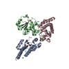
Mass: 35.453 Da / Num. of mol.: 2 / Source method: obtained synthetically / Formula: Cl
#3: Chemical ChemComp-FMT / FORMIC ACID / Formic acid


Mass: 46.025 Da / Num. of mol.: 3 / Source method: obtained synthetically / Formula: CH2O2

-
Experimental details

-
Experiment

ExperimentMethod: X-RAY DIFFRACTION / Number of used crystals: 1

-
Sample preparation

CrystalDensity Matthews: 3.13 Å3/Da / Density % sol: 60.64 %
Crystal growTemperature: 293 K / Method: vapor diffusion, sitting drop
Details: 0.1M Bis-Tris (pH 6.25) and 0.8M magnesium formate dehydrate

-
Data collection

DiffractionMean temperature: 93 K
Diffraction sourceSource: SYNCHROTRON / Site: PAL/PLS / Beamline: 7A (6B, 6C1) / Wavelength: 1 Å
DetectorType: ADSC QUANTUM 270 / Detector: CCD / Date: Sep 14, 2017
RadiationProtocol: SINGLE WAVELENGTH / Monochromatic (M) / Laue (L): M / Scattering type: x-ray
Radiation wavelengthWavelength: 1 Å / Relative weight: 1
ReflectionResolution: 2.298→77.4 Å / Num. obs: 28515 / % possible obs: 95.2 % / Redundancy: 4.3 % / Net I/σ(I): 27
Reflection shellResolution: 2.298→2.34 Å

-
Processing

Software
NameVersionClassification
REFMAC5.8.0158refinement
HKL-2000data reduction
HKL-2000data scaling
PHENIXphasing
RefinementMethod to determine structure: MOLECULAR REPLACEMENT
Starting model: 5Z5A
Resolution: 2.3→77.4 Å / Cor.coef. Fo:Fc: 0.921 / Cor.coef. Fo:Fc free: 0.881 / SU B: 8.346 / SU ML: 0.193 / Cross valid method: THROUGHOUT / ESU R: 0.31 / ESU R Free: 0.243 / Details: HYDROGENS HAVE BEEN ADDED IN THE RIDING POSITIONS
RfactorNum. reflection% reflectionSelection details
Rfree0.26459 1374 5.1 %RANDOM
Rwork0.21923 ---
obs0.2216 25754 90.58 %-
Solvent computationIon probe radii: 0.8 Å / Shrinkage radii: 0.8 Å / VDW probe radii: 1.2 Å
Displacement parametersBiso mean: 37.539 Å2
Baniso -1Baniso -2Baniso -3
1--0.16 Å2-0.08 Å20 Å2
2---0.16 Å20 Å2
3---0.53 Å2
Refinement stepCycle: 1 / Resolution: 2.3→77.4 Å
ProteinNucleic acidLigandSolventTotal
Num. atoms3707 0 11 0 3718
Refine LS restraints
Refine-IDTypeDev idealDev ideal targetNumber
X-RAY DIFFRACTIONr_bond_refined_d0.0160.0193782
X-RAY DIFFRACTIONr_bond_other_d0.0020.023687
X-RAY DIFFRACTIONr_angle_refined_deg1.8681.9795096
X-RAY DIFFRACTIONr_angle_other_deg1.07738487
X-RAY DIFFRACTIONr_dihedral_angle_1_deg7.3845458
X-RAY DIFFRACTIONr_dihedral_angle_2_deg33.9922.67176
X-RAY DIFFRACTIONr_dihedral_angle_3_deg18.44915702
X-RAY DIFFRACTIONr_dihedral_angle_4_deg17.1651544
X-RAY DIFFRACTIONr_chiral_restr0.10.2559
X-RAY DIFFRACTIONr_gen_planes_refined0.0080.0214144
X-RAY DIFFRACTIONr_gen_planes_other0.0020.02800
X-RAY DIFFRACTIONr_nbd_refined
X-RAY DIFFRACTIONr_nbd_other
X-RAY DIFFRACTIONr_nbtor_refined
X-RAY DIFFRACTIONr_nbtor_other
X-RAY DIFFRACTIONr_xyhbond_nbd_refined
X-RAY DIFFRACTIONr_xyhbond_nbd_other
X-RAY DIFFRACTIONr_metal_ion_refined
X-RAY DIFFRACTIONr_metal_ion_other
X-RAY DIFFRACTIONr_symmetry_vdw_refined
X-RAY DIFFRACTIONr_symmetry_vdw_other
X-RAY DIFFRACTIONr_symmetry_hbond_refined
X-RAY DIFFRACTIONr_symmetry_hbond_other
X-RAY DIFFRACTIONr_symmetry_metal_ion_refined
X-RAY DIFFRACTIONr_symmetry_metal_ion_other
X-RAY DIFFRACTIONr_mcbond_it2.853.5751842
X-RAY DIFFRACTIONr_mcbond_other2.853.5751843
X-RAY DIFFRACTIONr_mcangle_it4.4455.3542296
X-RAY DIFFRACTIONr_mcangle_other4.4445.3562297
X-RAY DIFFRACTIONr_scbond_it3.2673.9821940
X-RAY DIFFRACTIONr_scbond_other3.2673.9881935
X-RAY DIFFRACTIONr_scangle_it
X-RAY DIFFRACTIONr_scangle_other5.225.8172801
X-RAY DIFFRACTIONr_long_range_B_refined6.95240.7374078
X-RAY DIFFRACTIONr_long_range_B_other6.95140.7534079
X-RAY DIFFRACTIONr_rigid_bond_restr
X-RAY DIFFRACTIONr_sphericity_free
X-RAY DIFFRACTIONr_sphericity_bonded
LS refinement shellResolution: 2.298→2.358 Å / Total num. of bins used: 20
RfactorNum. reflection% reflection
Rfree0.401 60 -
Rwork0.286 1363 -
obs--64.27 %

+
About Yorodumi

-
News

-
Feb 9, 2022. New format data for meta-information of EMDB entries

New format data for meta-information of EMDB entries

  • Version 3 of the EMDB header file is now the official format.
  • The previous official version 1.9 will be removed from the archive.

Related info.:EMDB header

External links:wwPDB to switch to version 3 of the EMDB data model

-
Aug 12, 2020. Covid-19 info

Covid-19 info

URL: https://pdbj.org/emnavi/covid19.php

New page: Covid-19 featured information page in EM Navigator.

Related info.:Covid-19 info / Mar 5, 2020. Novel coronavirus structure data

+
Mar 5, 2020. Novel coronavirus structure data

Novel coronavirus structure data

Related info.:Yorodumi Speices / Aug 12, 2020. Covid-19 info

External links:COVID-19 featured content - PDBj / Molecule of the Month (242):Coronavirus Proteases

+
Jan 31, 2019. EMDB accession codes are about to change! (news from PDBe EMDB page)

EMDB accession codes are about to change! (news from PDBe EMDB page)

  • The allocation of 4 digits for EMDB accession codes will soon come to an end. Whilst these codes will remain in use, new EMDB accession codes will include an additional digit and will expand incrementally as the available range of codes is exhausted. The current 4-digit format prefixed with “EMD-” (i.e. EMD-XXXX) will advance to a 5-digit format (i.e. EMD-XXXXX), and so on. It is currently estimated that the 4-digit codes will be depleted around Spring 2019, at which point the 5-digit format will come into force.
  • The EM Navigator/Yorodumi systems omit the EMD- prefix.

Related info.:Q: What is EMD? / ID/Accession-code notation in Yorodumi/EM Navigator

External links:EMDB Accession Codes are Changing Soon! / Contact to PDBj

+
Jul 12, 2017. Major update of PDB

Major update of PDB

  • wwPDB released updated PDB data conforming to the new PDBx/mmCIF dictionary.
  • This is a major update changing the version number from 4 to 5, and with Remediation, in which all the entries are updated.
  • In this update, many items about electron microscopy experimental information are reorganized (e.g. em_software).
  • Now, EM Navigator and Yorodumi are based on the updated data.

External links:wwPDB Remediation / Enriched Model Files Conforming to OneDep Data Standards Now Available in the PDB FTP Archive

-
Yorodumi

Thousand views of thousand structures

  • Yorodumi is a browser for structure data from EMDB, PDB, SASBDB, etc.
  • This page is also the successor to EM Navigator detail page, and also detail information page/front-end page for Omokage search.
  • The word "yorodu" (or yorozu) is an old Japanese word meaning "ten thousand". "mi" (miru) is to see.

Related info.:EMDB / PDB / SASBDB / Comparison of 3 databanks / Yorodumi Search / Aug 31, 2016. New EM Navigator & Yorodumi / Yorodumi Papers / Jmol/JSmol / Function and homology information / Changes in new EM Navigator and Yorodumi

Read more