- PDB-5z2n: Structure of Orp1L N-terminal Domain -
+
Open data
ID or keywords:
Loading...
-
Basic information
Entry
Database: PDB / ID: 5z2n
Title
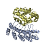
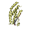
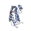
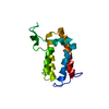
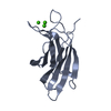
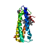
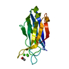
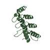
Structure of Orp1L N-terminal Domain
Components
Oxysterol-binding protein-related protein 1
Keywords
ENDOCYTOSIS / GTPase / Effector / Complex
Function / homology
Function and homology information
lysosome to ER cholesterol transport / sterol sensor activity / endoplasmic reticulum-endosome membrane contact site / Synthesis of bile acids and bile salts / organelle membrane contact site / perinuclear endoplasmic reticulum / MHC class II antigen presentation / cholesterol binding / cholesterol homeostasis / late endosome ...lysosome to ER cholesterol transport / sterol sensor activity / endoplasmic reticulum-endosome membrane contact site / Synthesis of bile acids and bile salts / organelle membrane contact site / perinuclear endoplasmic reticulum / MHC class II antigen presentation / cholesterol binding / cholesterol homeostasis / late endosome / endosome / plasma membrane / cytosol Similarity search - Function
Oxysterol-binding protein / Oxysterol-binding protein, conserved site / Oxysterol-binding protein superfamily / Oxysterol-binding protein / Oxysterol-binding protein family signature. / Ankyrin repeat-containing domain / PH domain profile. / Pleckstrin homology domain. / Pleckstrin homology domain / Ankyrin repeat profile. ...Oxysterol-binding protein / Oxysterol-binding protein, conserved site / Oxysterol-binding protein superfamily / Oxysterol-binding protein / Oxysterol-binding protein family signature. / Ankyrin repeat-containing domain / PH domain profile. / Pleckstrin homology domain. / Pleckstrin homology domain / Ankyrin repeat profile. / Ankyrin repeats (3 copies) / Ankyrin repeat region circular profile. / ankyrin repeats / Ankyrin repeat / Ankyrin repeat-containing domain superfamily / Serine Threonine Protein Phosphatase 5, Tetratricopeptide repeat / Alpha Horseshoe / PH-like domain superfamily / Mainly Alpha Similarity search - Domain/homology
In the structure databanks used in Yorodumi, some data are registered as the other names, "COVID-19 virus" and "2019-nCoV". Here are the details of the virus and the list of structure data.
Jan 31, 2019. EMDB accession codes are about to change! (news from PDBe EMDB page)
EMDB accession codes are about to change! (news from PDBe EMDB page)
The allocation of 4 digits for EMDB accession codes will soon come to an end. Whilst these codes will remain in use, new EMDB accession codes will include an additional digit and will expand incrementally as the available range of codes is exhausted. The current 4-digit format prefixed with “EMD-” (i.e. EMD-XXXX) will advance to a 5-digit format (i.e. EMD-XXXXX), and so on. It is currently estimated that the 4-digit codes will be depleted around Spring 2019, at which point the 5-digit format will come into force.
The EM Navigator/Yorodumi systems omit the EMD- prefix.
Related info.:Q: What is EMD? / ID/Accession-code notation in Yorodumi/EM Navigator
Yorodumi is a browser for structure data from EMDB, PDB, SASBDB, etc.
This page is also the successor to EM Navigator detail page, and also detail information page/front-end page for Omokage search.
The word "yorodu" (or yorozu) is an old Japanese word meaning "ten thousand". "mi" (miru) is to see.
Related info.:EMDB / PDB / SASBDB / Comparison of 3 databanks / Yorodumi Search / Aug 31, 2016. New EM Navigator & Yorodumi / Yorodumi Papers / Jmol/JSmol / Function and homology information / Changes in new EM Navigator and Yorodumi
 Movie
Movie Controller


 Open data
 Basic information
 Components
 Keywords
 Function and homology information
 X-RAY DIFFRACTION /
  Authors
 Citation
 Structure visualization
 Downloads & links
 Other downloads


 PDBj
PDBj


 Assembly


 Mass: 18.015 Da / Num. of mol.: 37 / Source method: isolated from a natural source / Formula: H2O
 Sample preparation
 / Beamline: X10SA / Wavelength: 1 Å
 Processing