 Movie
Movie Controller


+ Open data

- Basic information
| Entry | Database: PDB / ID: 5z09 | ||||||
|---|---|---|---|---|---|---|---|
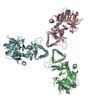
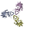
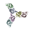
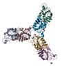
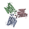
| Title | ST0452(Y97N)-UTP binding form | ||||||
 Components Components | Dual sugar-1-phosphate nucleotidylyltransferase | ||||||
 Keywords Keywords | SUGAR BINDING PROTEIN / UTP binding | ||||||
| Function / homology |  Function and homology information Function and homology informationgalactosamine-1-phosphate N-acetyltransferase / UDP-N-acetylgalactosamine diphosphorylase / UDP-N-acetylgalactosamine diphosphorylase activity / glucosamine-1-phosphate N-acetyltransferase / glucosamine-1-phosphate N-acetyltransferase activity / UTP-glucose-1-phosphate uridylyltransferase / UTP:glucose-1-phosphate uridylyltransferase activity / glucose-1-phosphate thymidylyltransferase / glucose-1-phosphate thymidylyltransferase activity / UDP-N-acetylglucosamine diphosphorylase ...galactosamine-1-phosphate N-acetyltransferase / UDP-N-acetylgalactosamine diphosphorylase / UDP-N-acetylgalactosamine diphosphorylase activity / glucosamine-1-phosphate N-acetyltransferase / glucosamine-1-phosphate N-acetyltransferase activity / UTP-glucose-1-phosphate uridylyltransferase / UTP:glucose-1-phosphate uridylyltransferase activity / glucose-1-phosphate thymidylyltransferase / glucose-1-phosphate thymidylyltransferase activity / UDP-N-acetylglucosamine diphosphorylase / UDP-N-acetylglucosamine diphosphorylase activity / UDP-N-acetylglucosamine biosynthetic process / nucleotide binding Similarity search - Function | ||||||
| Biological species |   Sulfolobus tokodaii (archaea) Sulfolobus tokodaii (archaea) | ||||||
| Method |  X-RAY DIFFRACTION / SYNCHROTRON / MOLECULAR REPLACEMENT / Resolution: 2.91 Å X-RAY DIFFRACTION / SYNCHROTRON / MOLECULAR REPLACEMENT / Resolution: 2.91 Å | ||||||
 Authors Authors | Honda, Y. / Nakano, S. / Ito, S. / Dadashipour, M. / Zhang, Z. / Kawarabayasi, Y. | ||||||
 Citation Citation | Journal: Appl. Environ. Microbiol. / Year: 2018 Title: Improvement of ST0452N-Acetylglucosamine-1-Phosphate Uridyltransferase Activity by the Cooperative Effect of Two Single Mutations Identified through Structure-Based Protein Engineering Authors: Honda, Y. / Nakano, S. / Ito, S. / Dadashipour, M. / Zhang, Z. / Kawarabayasi, Y. | ||||||
| History |
|
- Structure visualization
| Structure viewer | Molecule: MolmilJmol/JSmol |
|---|
- Downloads & links
-Download
| PDBx/mmCIF format | 5z09.cif.gz | 458.5 KB | Display | PDBx/mmCIF format |
|---|---|---|---|---|
| PDB format | pdb5z09.ent.gz | 373.7 KB | Display | PDB format |
| PDBx/mmJSON format | 5z09.json.gz | Tree view | PDBx/mmJSON format | |
| Others |  Other downloads Other downloads |
-Validation report
| Arichive directory | https://data.pdbj.org/pub/pdb/validation_reports/z0/5z09ftp://data.pdbj.org/pub/pdb/validation_reports/z0/5z09 | HTTPS FTP |
|---|
-Related structure data
| Related structure data |  5z0aC 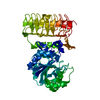 2ggoS S: Starting model for refinement C: citing same article ( |
|---|---|
| Similar structure data |


-Links
 PDBj
PDBj


- Assembly
| Deposited unit | 
| |||||||||||||||||||||||||||||||||||||||||||||||||||||||||||||||||||||||||||||||||||||||||||||||||||||||||||||||||||||||||||||||||||||||||||||||||||||||||||||||||||||||||||||||||||||||||||||||||||||||||||||||||||||||||||||||||||||||||
|---|---|---|---|---|---|---|---|---|---|---|---|---|---|---|---|---|---|---|---|---|---|---|---|---|---|---|---|---|---|---|---|---|---|---|---|---|---|---|---|---|---|---|---|---|---|---|---|---|---|---|---|---|---|---|---|---|---|---|---|---|---|---|---|---|---|---|---|---|---|---|---|---|---|---|---|---|---|---|---|---|---|---|---|---|---|---|---|---|---|---|---|---|---|---|---|---|---|---|---|---|---|---|---|---|---|---|---|---|---|---|---|---|---|---|---|---|---|---|---|---|---|---|---|---|---|---|---|---|---|---|---|---|---|---|---|---|---|---|---|---|---|---|---|---|---|---|---|---|---|---|---|---|---|---|---|---|---|---|---|---|---|---|---|---|---|---|---|---|---|---|---|---|---|---|---|---|---|---|---|---|---|---|---|---|---|---|---|---|---|---|---|---|---|---|---|---|---|---|---|---|---|---|---|---|---|---|---|---|---|---|---|---|---|---|---|---|---|---|---|---|---|---|---|---|---|---|---|---|---|---|---|---|---|---|
| 1 | 
| |||||||||||||||||||||||||||||||||||||||||||||||||||||||||||||||||||||||||||||||||||||||||||||||||||||||||||||||||||||||||||||||||||||||||||||||||||||||||||||||||||||||||||||||||||||||||||||||||||||||||||||||||||||||||||||||||||||||||
| 2 | 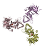
| |||||||||||||||||||||||||||||||||||||||||||||||||||||||||||||||||||||||||||||||||||||||||||||||||||||||||||||||||||||||||||||||||||||||||||||||||||||||||||||||||||||||||||||||||||||||||||||||||||||||||||||||||||||||||||||||||||||||||
| Unit cell |
| |||||||||||||||||||||||||||||||||||||||||||||||||||||||||||||||||||||||||||||||||||||||||||||||||||||||||||||||||||||||||||||||||||||||||||||||||||||||||||||||||||||||||||||||||||||||||||||||||||||||||||||||||||||||||||||||||||||||||
| Noncrystallographic symmetry (NCS) | NCS domain:
NCS domain segments: Component-ID: _ / Beg auth comp-ID: MET / Beg label comp-ID: MET / End auth comp-ID: VAL / End label comp-ID: VAL / Refine code: _ / Auth seq-ID: 1 - 401 / Label seq-ID: 1 - 401
NCS ensembles :
|
-Components
| #1: Protein | Mass: 45595.430 Da / Num. of mol.: 6 Source method: isolated from a genetically manipulated source Source: (gene. exp.) Sulfolobus tokodaii (strain DSM 16993 / JCM 10545 / NBRC 100140 / 7) (archaea)Strain: DSM 16993 / JCM 10545 / NBRC 100140 / 7 / Gene: ST0452, STK_04520 / Production host:  References: UniProt: Q975F9, Transferases; Transferring phosphorus-containing groups; Nucleotidyltransferases #2: Chemical | ChemComp-UTP /   Mass: 484.141 Da / Num. of mol.: 4 / Source method: obtained synthetically / Formula: C9H15N2O15P3 / Comment: UTP*YM Mass: 484.141 Da / Num. of mol.: 4 / Source method: obtained synthetically / Formula: C9H15N2O15P3 / Comment: UTP*YMHas protein modification | Y | |
|---|
-Experimental details
-Experiment
| Experiment | Method: X-RAY DIFFRACTION / Number of used crystals: 1 |
|---|
- Sample preparation
| Crystal | Density Matthews: 2.6 Å3/Da / Density % sol: 52.7 % |
|---|---|
| Crystal grow | Temperature: 295 K / Method: vapor diffusion, sitting drop Details: 20%(w/v) PEG3350, 0.2M potassium citrate tribasic monohydrate |
-Data collection
| Diffraction | Mean temperature: 100 K |
|---|---|
| Diffraction source | Source: SYNCHROTRON / Site: Photon Factory  / Beamline: BL-5A / Wavelength: 1 Å / Beamline: BL-5A / Wavelength: 1 Å |
| Detector | Type: DECTRIS PILATUS 2M-F / Detector: PIXEL / Date: Dec 5, 2016 |
| Radiation | Protocol: SINGLE WAVELENGTH / Monochromatic (M) / Laue (L): M / Scattering type: x-ray |
| Radiation wavelength | Wavelength: 1 Å / Relative weight: 1 |
| Reflection | Resolution: 2.9→153 Å / Num. obs: 61139 / % possible obs: 99 % / Redundancy: 6.8 % / Rmerge(I) obs: 0.092 / Net I/σ(I): 16.6 |
| Reflection shell | Resolution: 2.9→2.98 Å / Rmerge(I) obs: 0.513 / Num. unique obs: 3018 |
- Processing
| Software |
| ||||||||||||||||||||||||||||||||||||||||||||||||||||||||||||||||||||||||||||||||||||||||||||||||||||||||||||||||||||||||||||||||||||||||||||||||||||||||||||||||||||||||||||||||||||||
|---|---|---|---|---|---|---|---|---|---|---|---|---|---|---|---|---|---|---|---|---|---|---|---|---|---|---|---|---|---|---|---|---|---|---|---|---|---|---|---|---|---|---|---|---|---|---|---|---|---|---|---|---|---|---|---|---|---|---|---|---|---|---|---|---|---|---|---|---|---|---|---|---|---|---|---|---|---|---|---|---|---|---|---|---|---|---|---|---|---|---|---|---|---|---|---|---|---|---|---|---|---|---|---|---|---|---|---|---|---|---|---|---|---|---|---|---|---|---|---|---|---|---|---|---|---|---|---|---|---|---|---|---|---|---|---|---|---|---|---|---|---|---|---|---|---|---|---|---|---|---|---|---|---|---|---|---|---|---|---|---|---|---|---|---|---|---|---|---|---|---|---|---|---|---|---|---|---|---|---|---|---|---|---|
| Refinement | Method to determine structure: MOLECULAR REPLACEMENT Starting model: 2GGO Resolution: 2.91→152.76 Å / Cor.coef. Fo:Fc: 0.932 / Cor.coef. Fo:Fc free: 0.899 / SU B: 19.996 / SU ML: 0.365 / Cross valid method: THROUGHOUT / ESU R Free: 0.445 / Details: HYDROGENS HAVE BEEN ADDED IN THE RIDING POSITIONS
| ||||||||||||||||||||||||||||||||||||||||||||||||||||||||||||||||||||||||||||||||||||||||||||||||||||||||||||||||||||||||||||||||||||||||||||||||||||||||||||||||||||||||||||||||||||||
| Solvent computation | Ion probe radii: 0.8 Å / Shrinkage radii: 0.8 Å / VDW probe radii: 1.2 Å | ||||||||||||||||||||||||||||||||||||||||||||||||||||||||||||||||||||||||||||||||||||||||||||||||||||||||||||||||||||||||||||||||||||||||||||||||||||||||||||||||||||||||||||||||||||||
| Displacement parameters | Biso mean: 83.069 Å2
| ||||||||||||||||||||||||||||||||||||||||||||||||||||||||||||||||||||||||||||||||||||||||||||||||||||||||||||||||||||||||||||||||||||||||||||||||||||||||||||||||||||||||||||||||||||||
| Refinement step | Cycle: 1 / Resolution: 2.91→152.76 Å
| ||||||||||||||||||||||||||||||||||||||||||||||||||||||||||||||||||||||||||||||||||||||||||||||||||||||||||||||||||||||||||||||||||||||||||||||||||||||||||||||||||||||||||||||||||||||
| Refine LS restraints |
|