 Movie
Movie Controller

[English] 日本語
 Yorodumi
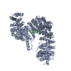
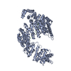
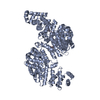
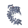
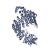
- PDB-5yvh: Crystal structure of Karyopherin beta2 in complex with FUS(371-526) -
+ Open data

- Basic information
| Entry | Database: PDB / ID: 5yvh | ||||||
|---|---|---|---|---|---|---|---|
| Title | Crystal structure of Karyopherin beta2 in complex with FUS(371-526) | ||||||
 Components Components |
| ||||||
 Keywords Keywords | PROTEIN TRANSPORT/RNA BINDING PROTEIN / Importin family / TRANSPORT PROTEIN / PROTEIN TRANSPORT-RNA BINDING PROTEIN complex | ||||||
| Function / homology |  Function and homology information Function and homology informationTristetraprolin (TTP, ZFP36) binds and destabilizes mRNA / Intraflagellar transport / membraneless organelle assembly / Postmitotic nuclear pore complex (NPC) reformation / nuclear import signal receptor activity / nuclear localization sequence binding / mRNA stabilization / regulation of RNA splicing / Processing of Capped Intron-Containing Pre-mRNA / postsynaptic cytosol ...Tristetraprolin (TTP, ZFP36) binds and destabilizes mRNA / Intraflagellar transport / membraneless organelle assembly / Postmitotic nuclear pore complex (NPC) reformation / nuclear import signal receptor activity / nuclear localization sequence binding / mRNA stabilization / regulation of RNA splicing / Processing of Capped Intron-Containing Pre-mRNA / postsynaptic cytosol / positive regulation of double-strand break repair via homologous recombination / presynaptic cytosol / mRNA Splicing - Major Pathway / RNA splicing / transcription coregulator activity / mRNA 3'-UTR binding / molecular condensate scaffold activity / protein homooligomerization / GABA-ergic synapse / small GTPase binding / protein import into nucleus / amyloid fibril formation / transcription coactivator activity / cilium / chromatin binding / regulation of transcription by RNA polymerase II / regulation of DNA-templated transcription / nucleolus / glutamatergic synapse / DNA binding / RNA binding / extracellular exosome / zinc ion binding / nucleoplasm / identical protein binding / nucleus / cytoplasm / cytosol Similarity search - Function | ||||||
| Biological species |  Homo sapiens (human) Homo sapiens (human) | ||||||
| Method |  X-RAY DIFFRACTION / SYNCHROTRON / MOLECULAR REPLACEMENT / Resolution: 3.15 Å X-RAY DIFFRACTION / SYNCHROTRON / MOLECULAR REPLACEMENT / Resolution: 3.15 Å | ||||||
 Authors Authors | Yoshizawa, T. / Fung, H.Y.J. / Chook, Y.M. | ||||||
 Citation Citation | Journal: Cell / Year: 2018 Title: Nuclear Import Receptor Inhibits Phase Separation of FUS through Binding to Multiple Sites. Authors: Yoshizawa, T. / Ali, R. / Jiou, J. / Fung, H.Y.J. / Burke, K.A. / Kim, S.J. / Lin, Y. / Peeples, W.B. / Saltzberg, D. / Soniat, M. / Baumhardt, J.M. / Oldenbourg, R. / Sali, A. / Fawzi, N.L. ...Authors: Yoshizawa, T. / Ali, R. / Jiou, J. / Fung, H.Y.J. / Burke, K.A. / Kim, S.J. / Lin, Y. / Peeples, W.B. / Saltzberg, D. / Soniat, M. / Baumhardt, J.M. / Oldenbourg, R. / Sali, A. / Fawzi, N.L. / Rosen, M.K. / Chook, Y.M. | ||||||
| History |
|
- Structure visualization
| Structure viewer | Molecule: MolmilJmol/JSmol |
|---|
- Downloads & links
-Download
| PDBx/mmCIF format | 5yvh.cif.gz | 346.8 KB | Display | PDBx/mmCIF format |
|---|---|---|---|---|
| PDB format | pdb5yvh.ent.gz | 281.2 KB | Display | PDB format |
| PDBx/mmJSON format | 5yvh.json.gz | Tree view | PDBx/mmJSON format | |
| Others |  Other downloads Other downloads |
-Validation report
| Arichive directory | https://data.pdbj.org/pub/pdb/validation_reports/yv/5yvhftp://data.pdbj.org/pub/pdb/validation_reports/yv/5yvh | HTTPS FTP |
|---|
-Related structure data
| Related structure data | 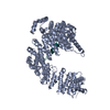 5yvgC  5yviC  4fddS S: Starting model for refinement C: citing same article ( |
|---|---|
| Similar structure data |


-Links
 PDBj
PDBj


- Assembly
| Deposited unit | 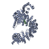
| ||||||||
|---|---|---|---|---|---|---|---|---|---|
| 1 |
| ||||||||
| Unit cell |
|
-Components
| #1: Protein | Mass: 98325.352 Da / Num. of mol.: 1 / Fragment: TRUNCATED RESIDUES 345-375 REPLACED WITH GGSGGSG Source method: isolated from a genetically manipulated source Source: (gene. exp.) Homo sapiens (human) / Gene: TNPO1, KPNB2, MIP1, TRN / Production host:  |
|---|---|
| #2: Protein | Mass: 15983.178 Da / Num. of mol.: 1 / Fragment: Arg/Gly-rich domain Source method: isolated from a genetically manipulated source Source: (gene. exp.) Homo sapiens (human) / Gene: FUS, TLS / Production host: |
-Experimental details
-Experiment
| Experiment | Method: X-RAY DIFFRACTION / Number of used crystals: 1 |
|---|
- Sample preparation
| Crystal | Density Matthews: 3.01 Å3/Da / Density % sol: 59.07 % |
|---|---|
| Crystal grow | Temperature: 293 K / Method: vapor diffusion, hanging drop / Details: 0.8M Succinic acid pH7.0 |
-Data collection
| Diffraction | Mean temperature: 93 K |
|---|---|
| Diffraction source | Source: SYNCHROTRON / Site: APS  / Beamline: 19-ID / Wavelength: 0.97926 Å / Beamline: 19-ID / Wavelength: 0.97926 Å |
| Detector | Type: ADSC QUANTUM 315r / Detector: CCD / Date: Feb 14, 2013 |
| Radiation | Protocol: SINGLE WAVELENGTH / Monochromatic (M) / Laue (L): M / Scattering type: x-ray |
| Radiation wavelength | Wavelength: 0.97926 Å / Relative weight: 1 |
| Reflection | Resolution: 3.15→50 Å / Num. obs: 25064 / % possible obs: 99.6 % / Redundancy: 5.4 % / Rpim(I) all: 0.051 / Net I/av σ(I): 11.99 / Net I/σ(I): 5.4 |
| Reflection shell | Resolution: 3.15→3.2 Å / Mean I/σ(I) obs: 1.34 / CC1/2: 0.68 / Rpim(I) all: 0.533 |
- Processing
| Software |
| |||||||||||||||||||||||||||||||||||||||||||||||||||||||||||||||
|---|---|---|---|---|---|---|---|---|---|---|---|---|---|---|---|---|---|---|---|---|---|---|---|---|---|---|---|---|---|---|---|---|---|---|---|---|---|---|---|---|---|---|---|---|---|---|---|---|---|---|---|---|---|---|---|---|---|---|---|---|---|---|---|---|
| Refinement | Method to determine structure: MOLECULAR REPLACEMENT Starting model: 4FDD Resolution: 3.15→41.355 Å / SU ML: 0.27 / Cross valid method: FREE R-VALUE / σ(F): 1.35 / Phase error: 26.49
| |||||||||||||||||||||||||||||||||||||||||||||||||||||||||||||||
| Solvent computation | Shrinkage radii: 0.9 Å / VDW probe radii: 1.11 Å | |||||||||||||||||||||||||||||||||||||||||||||||||||||||||||||||
| Refinement step | Cycle: LAST / Resolution: 3.15→41.355 Å
| |||||||||||||||||||||||||||||||||||||||||||||||||||||||||||||||
| Refine LS restraints |
| |||||||||||||||||||||||||||||||||||||||||||||||||||||||||||||||
| LS refinement shell |
| |||||||||||||||||||||||||||||||||||||||||||||||||||||||||||||||
| Refinement TLS params. | Method: refined / Origin x: 29.5799 Å / Origin y: 41.2374 Å / Origin z: 30.2869 Å
| |||||||||||||||||||||||||||||||||||||||||||||||||||||||||||||||
| Refinement TLS group |
|
