 Movie
Movie Controller

[English] 日本語
 Yorodumi
- PDB-5ycs: X-Ray Structure of Enoyl-Acyl Carrier Protein Reductase from Baci... -
+ Open data

- Basic information
| Entry | Database: PDB / ID: 5ycs | ||||||
|---|---|---|---|---|---|---|---|
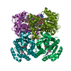
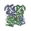
| Title | X-Ray Structure of Enoyl-Acyl Carrier Protein Reductase from Bacillus Anthracis with triclosan | ||||||
 Components Components | Enoyl-[acyl-carrier-protein] reductase [NADH] FabI | ||||||
 Keywords Keywords | OXIDOREDUCTASE / antibacterial / fabI | ||||||
| Function / homology |  Function and homology information Function and homology informationenoyl-[acyl-carrier-protein] reductase (NADPH) activity / enoyl-[acyl-carrier-protein] reductase (NADH) / fatty acid elongation / enoyl-[acyl-carrier-protein] reductase (NADH) activity / NADP+ binding / small molecule binding / catalytic complex / identical protein binding Similarity search - Function | ||||||
| Biological species |  | ||||||
| Method |  X-RAY DIFFRACTION / SYNCHROTRON / MOLECULAR REPLACEMENT / Resolution: 1.95 Å X-RAY DIFFRACTION / SYNCHROTRON / MOLECULAR REPLACEMENT / Resolution: 1.95 Å | ||||||
 Authors Authors | Kim, H.T. | ||||||
 Citation Citation | Journal: Biochem. Biophys. Res. Commun. / Year: 2017 Title: Structural insights into the dimer-tetramer transition of FabI from Bacillus anthracis Authors: Kim, H.T. / Kim, S. / Na, B.K. / Chung, J. / Hwang, E. / Hwang, K.Y. | ||||||
| History |
|
- Structure visualization
| Structure viewer | Molecule: MolmilJmol/JSmol |
|---|
- Downloads & links
-Download
| PDBx/mmCIF format | 5ycs.cif.gz | 225.2 KB | Display | PDBx/mmCIF format |
|---|---|---|---|---|
| PDB format | pdb5ycs.ent.gz | 180.5 KB | Display | PDB format |
| PDBx/mmJSON format | 5ycs.json.gz | Tree view | PDBx/mmJSON format | |
| Others |  Other downloads Other downloads |
-Validation report
| Arichive directory | https://data.pdbj.org/pub/pdb/validation_reports/yc/5ycsftp://data.pdbj.org/pub/pdb/validation_reports/yc/5ycs | HTTPS FTP |
|---|
-Related structure data
| Related structure data |  5ycrC  5ycvC  5ycxC  2qioS S: Starting model for refinement C: citing same article ( |
|---|---|
| Similar structure data |


-Links
 PDBj
PDBj

- Assembly
| Deposited unit | 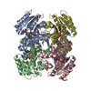
| ||||||||
|---|---|---|---|---|---|---|---|---|---|
| 1 |
| ||||||||
| Unit cell |
| ||||||||
| Components on special symmetry positions |
|
-Components
| #1: Protein | Mass: 27916.555 Da / Num. of mol.: 4 / Mutation: no Source method: isolated from a genetically manipulated source Details: NAD+, triclosan Source: (gene. exp.) Strain: ATCC 14579 / DSM 31 / JCM 2152 / NBRC 15305 / NCIMB 9373 / NRRL B-3711 Gene: fabI, BC_1216 Production host: References: UniProt: Q81GI3, enoyl-[acyl-carrier-protein] reductase (NADH) #2: Chemical | ChemComp-NAD /   Mass: 663.425 Da / Num. of mol.: 4 / Source method: obtained synthetically / Formula: C21H27N7O14P2 / Comment: NAD*YM Mass: 663.425 Da / Num. of mol.: 4 / Source method: obtained synthetically / Formula: C21H27N7O14P2 / Comment: NAD*YM#3: Chemical | ChemComp-TCL /   Mass: 289.542 Da / Num. of mol.: 4 / Source method: obtained synthetically / Formula: C12H7Cl3O2 / Comment: antifungal, antibiotic, detergent*YM Mass: 289.542 Da / Num. of mol.: 4 / Source method: obtained synthetically / Formula: C12H7Cl3O2 / Comment: antifungal, antibiotic, detergent*YM#4: Chemical |   Mass: 96.063 Da / Num. of mol.: 2 / Source method: obtained synthetically / Formula: SO4 Mass: 96.063 Da / Num. of mol.: 2 / Source method: obtained synthetically / Formula: SO4#5: Water | ChemComp-HOH / |  Mass: 18.015 Da / Num. of mol.: 689 / Source method: isolated from a natural source / Formula: H2O Mass: 18.015 Da / Num. of mol.: 689 / Source method: isolated from a natural source / Formula: H2O |
|---|
-Experimental details
-Experiment
| Experiment | Method: X-RAY DIFFRACTION / Number of used crystals: 1 |
|---|
- Sample preparation
| Crystal | Density Matthews: 3.82 Å3/Da / Density % sol: 67.77 % |
|---|---|
| Crystal grow | Temperature: 295 K / Method: vapor diffusion, hanging drop / pH: 4.6 / Details: 0.1M Na acetate, pH 4.6, 2M AMS |
-Data collection
| Diffraction | Mean temperature: 100 K |
|---|---|
| Diffraction source | Source: SYNCHROTRON / Site: PAL/PLS  / Beamline: 5C (4A) / Wavelength: 1 Å / Beamline: 5C (4A) / Wavelength: 1 Å |
| Detector | Type: ADSC QUANTUM 210r / Detector: CCD / Date: Oct 15, 2016 |
| Radiation | Protocol: SINGLE WAVELENGTH / Monochromatic (M) / Laue (L): M / Scattering type: x-ray |
| Radiation wavelength | Wavelength: 1 Å / Relative weight: 1 |
| Reflection | Resolution: 1.95→50 Å / Num. obs: 124185 / % possible obs: 99.8 % / Redundancy: 7.9 % / Rmerge(I) obs: 0.101 / Rpim(I) all: 0.035 / Rsym value: 0.086 / Net I/σ(I): 29 |
| Reflection shell | Resolution: 1.95→1.98 Å / Redundancy: 6.1 % / Rmerge(I) obs: 0.361 / Mean I/σ(I) obs: 3.5 / Num. unique obs: 6104 / CC1/2: 0.86 / Rpim(I) all: 0.151 / Rsym value: 0.475 / Χ2: 0.639 / % possible all: 99.5 |
- Processing
| Software |
| |||||||||||||||||||||||||||||||||||||||||||||||||||||||||||||||||||||||||||||||||||||||||||||||||||||||||
|---|---|---|---|---|---|---|---|---|---|---|---|---|---|---|---|---|---|---|---|---|---|---|---|---|---|---|---|---|---|---|---|---|---|---|---|---|---|---|---|---|---|---|---|---|---|---|---|---|---|---|---|---|---|---|---|---|---|---|---|---|---|---|---|---|---|---|---|---|---|---|---|---|---|---|---|---|---|---|---|---|---|---|---|---|---|---|---|---|---|---|---|---|---|---|---|---|---|---|---|---|---|---|---|---|---|---|
| Refinement | Method to determine structure: MOLECULAR REPLACEMENT Starting model: 2QIO Resolution: 1.95→36.595 Å / SU ML: 0.18 / Cross valid method: FREE R-VALUE / σ(F): 1.43 / Phase error: 17.49
| |||||||||||||||||||||||||||||||||||||||||||||||||||||||||||||||||||||||||||||||||||||||||||||||||||||||||
| Solvent computation | Shrinkage radii: 0.9 Å / VDW probe radii: 1.11 Å | |||||||||||||||||||||||||||||||||||||||||||||||||||||||||||||||||||||||||||||||||||||||||||||||||||||||||
| Refinement step | Cycle: LAST / Resolution: 1.95→36.595 Å
| |||||||||||||||||||||||||||||||||||||||||||||||||||||||||||||||||||||||||||||||||||||||||||||||||||||||||
| Refine LS restraints |
| |||||||||||||||||||||||||||||||||||||||||||||||||||||||||||||||||||||||||||||||||||||||||||||||||||||||||
| LS refinement shell |
|
