 Movie
Movie Controller


+ Open data

- Basic information
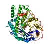
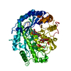
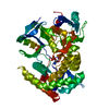
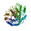
| Entry | Database: PDB / ID: 5y50 | ||||||
|---|---|---|---|---|---|---|---|
| Title | Crystal structure of eukaryotic MATE transporter AtDTX14 | ||||||
 Components Components | Protein DETOXIFICATION 14 | ||||||
 Keywords Keywords | MEMBRANE PROTEIN / alpha helical | ||||||
| Function / homology |  Function and homology information Function and homology informationplant-type vacuole / xenobiotic detoxification by transmembrane export across the plasma membrane / antiporter activity / xenobiotic transmembrane transporter activity / xenobiotic transport / membrane Similarity search - Function | ||||||
| Biological species |  | ||||||
| Method |  X-RAY DIFFRACTION / SYNCHROTRON / MOLECULAR REPLACEMENT / Resolution: 2.6 Å X-RAY DIFFRACTION / SYNCHROTRON / MOLECULAR REPLACEMENT / Resolution: 2.6 Å | ||||||
| Model details | a receptor | ||||||
 Authors Authors | Miyauchi, H. / Kusakizako, T. / Nishizawa, T. / Ishitani, R. / Nureki, O. | ||||||
 Citation Citation | Journal: Nat Commun / Year: 2017 Title: Structural basis for xenobiotic extrusion by eukaryotic MATE transporter Authors: Miyauchi, H. / Moriyama, S. / Kusakizako, T. / Kumazaki, K. / Nakane, T. / Yamashita, K. / Hirata, K. / Dohmae, N. / Nishizawa, T. / Ito, K. / Miyaji, T. / Moriyama, Y. / Ishitani, R. / Nureki, O. | ||||||
| History |
|
- Structure visualization
| Structure viewer | Molecule: MolmilJmol/JSmol |
|---|
- Downloads & links
-Download
| PDBx/mmCIF format | 5y50.cif.gz | 96.7 KB | Display | PDBx/mmCIF format |
|---|---|---|---|---|
| PDB format | pdb5y50.ent.gz | 72.1 KB | Display | PDB format |
| PDBx/mmJSON format | 5y50.json.gz | Tree view | PDBx/mmJSON format | |
| Others |  Other downloads Other downloads |
-Validation report
| Arichive directory | https://data.pdbj.org/pub/pdb/validation_reports/y5/5y50ftp://data.pdbj.org/pub/pdb/validation_reports/y5/5y50 | HTTPS FTP |
|---|
-Related structure data
| Related structure data |  3mktS S: Starting model for refinement |
|---|---|
| Similar structure data |


-Links
 PDBj
PDBj- Assembly
| Deposited unit | 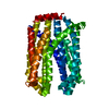
| ||||||||
|---|---|---|---|---|---|---|---|---|---|
| 1 |
| ||||||||
| Unit cell |
|
-Components
| #1: Protein | Mass: 49544.004 Da / Num. of mol.: 1 / Fragment: UNP residues 20-473 / Mutation: P36A Source method: isolated from a genetically manipulated source Source: (gene. exp.)   Spodoptera frugiperda (fall armyworm) / References: UniProt: Q9C994 Spodoptera frugiperda (fall armyworm) / References: UniProt: Q9C994 |
|---|
-Experimental details
-Experiment
| Experiment | Method: X-RAY DIFFRACTION / Number of used crystals: 1 |
|---|
- Sample preparation
| Crystal | Density Matthews: 2.69 Å3/Da / Density % sol: 54.31 % |
|---|---|
| Crystal grow | Temperature: 293 K / Method: lipidic cubic phase / pH: 5 Details: Na-citrate, MgSO4, NaK-tartrate-tetrahydrate, PEG550MME |
-Data collection
| Diffraction | Mean temperature: 100 K | ||||||||||||||||||||||||||||||||||||||||||||||||||||||||||||||||||||||||||||||||
|---|---|---|---|---|---|---|---|---|---|---|---|---|---|---|---|---|---|---|---|---|---|---|---|---|---|---|---|---|---|---|---|---|---|---|---|---|---|---|---|---|---|---|---|---|---|---|---|---|---|---|---|---|---|---|---|---|---|---|---|---|---|---|---|---|---|---|---|---|---|---|---|---|---|---|---|---|---|---|---|---|---|
| Diffraction source | Source: SYNCHROTRON / Site: SPring-8  / Beamline: BL32XU / Wavelength: 1 Å / Beamline: BL32XU / Wavelength: 1 Å | ||||||||||||||||||||||||||||||||||||||||||||||||||||||||||||||||||||||||||||||||
| Detector | Type: RAYONIX MX225-HS / Detector: CCD / Date: Oct 9, 2015 | ||||||||||||||||||||||||||||||||||||||||||||||||||||||||||||||||||||||||||||||||
| Radiation | Protocol: SINGLE WAVELENGTH / Monochromatic (M) / Laue (L): M / Scattering type: x-ray | ||||||||||||||||||||||||||||||||||||||||||||||||||||||||||||||||||||||||||||||||
| Radiation wavelength | Wavelength: 1 Å / Relative weight: 1 | ||||||||||||||||||||||||||||||||||||||||||||||||||||||||||||||||||||||||||||||||
| Reflection | Resolution: 2.6→48.345 Å / Num. obs: 16880 / % possible obs: 98.5 % / Observed criterion σ(I): -3 / Redundancy: 28.488 % / Biso Wilson estimate: 47.81 Å2 / CC1/2: 0.994 / Rmerge(I) obs: 0.576 / Rrim(I) all: 0.587 / Χ2: 1.197 / Net I/σ(I): 7.79 | ||||||||||||||||||||||||||||||||||||||||||||||||||||||||||||||||||||||||||||||||
| Reflection shell | Diffraction-ID: 1
|
- Processing
| Software |
| |||||||||||||||||||||||||||||||||||||||||||||||||
|---|---|---|---|---|---|---|---|---|---|---|---|---|---|---|---|---|---|---|---|---|---|---|---|---|---|---|---|---|---|---|---|---|---|---|---|---|---|---|---|---|---|---|---|---|---|---|---|---|---|---|
| Refinement | Method to determine structure: MOLECULAR REPLACEMENT Starting model: 3MKT Resolution: 2.6→48.345 Å / SU ML: 0.35 / Cross valid method: FREE R-VALUE / σ(F): 1.35 / Phase error: 29.87
| |||||||||||||||||||||||||||||||||||||||||||||||||
| Solvent computation | Shrinkage radii: 0.9 Å / VDW probe radii: 1.11 Å | |||||||||||||||||||||||||||||||||||||||||||||||||
| Displacement parameters | Biso max: 106.88 Å2 / Biso mean: 52.1982 Å2 / Biso min: 25.08 Å2 | |||||||||||||||||||||||||||||||||||||||||||||||||
| Refinement step | Cycle: final / Resolution: 2.6→48.345 Å
| |||||||||||||||||||||||||||||||||||||||||||||||||
| Refine LS restraints |
| |||||||||||||||||||||||||||||||||||||||||||||||||
| LS refinement shell | Refine-ID: X-RAY DIFFRACTION / Rfactor Rfree error: 0 / Total num. of bins used: 6
|
