 Movie
Movie Controller

[English] 日本語
 Yorodumi
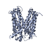
- PDB-3mkt: Structure of a Cation-bound Multidrug and Toxin Compound Extrusio... -
+ Open data

- Basic information
| Entry | Database: PDB / ID: 3mkt | ||||||
|---|---|---|---|---|---|---|---|
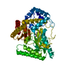
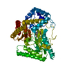
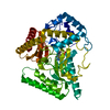
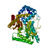
| Title | Structure of a Cation-bound Multidrug and Toxin Compound Extrusion (MATE) transporter | ||||||
 Components Components | Multi antimicrobial extrusion protein (Na(+)/drug antiporter) MATE-like MDR efflux pump | ||||||
 Keywords Keywords | TRANSPORT PROTEIN / MATE / multidrug transporter / cation-bound | ||||||
| Function / homology | :  Function and homology information Function and homology information | ||||||
| Biological species |   Vibrio cholerae (bacteria) Vibrio cholerae (bacteria) | ||||||
| Method |  X-RAY DIFFRACTION / SYNCHROTRON / SAD / Resolution: 3.65 Å X-RAY DIFFRACTION / SYNCHROTRON / SAD / Resolution: 3.65 Å | ||||||
 Authors Authors | He, X. / Szewczyk, P. / Karyakin, A. / Evin, M. / Hong, W.-X. / Zhang, Q. / Chang, G. | ||||||
 Citation Citation | Journal: Nature / Year: 2010 Title: Structure of a cation-bound multidrug and toxic compound extrusion transporter. Authors: He, X. / Szewczyk, P. / Karyakin, A. / Evin, M. / Hong, W.X. / Zhang, Q. / Chang, G. | ||||||
| History |
| ||||||
| Remark 650 | HELIX DETERMINATION METHOD: AUTHOR DETERMINED |
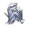
- Structure visualization
| Structure viewer | Molecule: MolmilJmol/JSmol |
|---|
- Downloads & links
-Download
| PDBx/mmCIF format | 3mkt.cif.gz | 167.1 KB | Display | PDBx/mmCIF format |
|---|---|---|---|---|
| PDB format | pdb3mkt.ent.gz | 134.6 KB | Display | PDB format |
| PDBx/mmJSON format | 3mkt.json.gz | Tree view | PDBx/mmJSON format | |
| Others |  Other downloads Other downloads |
-Validation report
| Arichive directory | https://data.pdbj.org/pub/pdb/validation_reports/mk/3mktftp://data.pdbj.org/pub/pdb/validation_reports/mk/3mkt | HTTPS FTP |
|---|
-Related structure data


-Links
 PDBj
PDBj- Assembly
| Deposited unit | 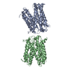
| ||||||||
|---|---|---|---|---|---|---|---|---|---|
| 1 | 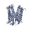
| ||||||||
| 2 | 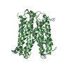
| ||||||||
| Unit cell |
|
-Components
| #1: Protein | Mass: 49761.133 Da / Num. of mol.: 2 Source method: isolated from a genetically manipulated source Source: (gene. exp.) Vibrio cholerae (bacteria) / Strain: N16961 / Gene: NorM / Plasmid: pET19b / Production host: |
|---|
-Experimental details
-Experiment
| Experiment | Method: X-RAY DIFFRACTION / Number of used crystals: 1 |
|---|
- Sample preparation
| Crystal | Density Matthews: 3.83 Å3/Da / Density % sol: 67.86 % |
|---|---|
| Crystal grow | Temperature: 295 K / Method: vapor diffusion, sitting drop / pH: 7.6 Details: 50mM Tris-HCl pH 7.2-8.6, 87 mM (NH4)2SO4, and 16-24% polyethylene glycol 250 dimethyl ether in D2O, VAPOR DIFFUSION, SITTING DROP, temperature 295K |
-Data collection
| Diffraction | Mean temperature: 100 K |
|---|---|
| Diffraction source | Source: SYNCHROTRON / Site: APS  / Beamline: 23-ID-B / Wavelength: 1.0093 Å / Beamline: 23-ID-B / Wavelength: 1.0093 Å |
| Detector | Type: MARMOSAIC 300 mm CCD / Detector: CCD / Date: Aug 13, 2009 |
| Radiation | Protocol: SINGLE WAVELENGTH / Monochromatic (M) / Laue (L): M / Scattering type: x-ray |
| Radiation wavelength | Wavelength: 1.0093 Å / Relative weight: 1 |
| Reflection | Resolution: 3.65→19.77 Å / Num. obs: 15364 / Biso Wilson estimate: 30.5 Å2 |
- Processing
| Software |
| ||||||||||||||||||||||||||||||||||||
|---|---|---|---|---|---|---|---|---|---|---|---|---|---|---|---|---|---|---|---|---|---|---|---|---|---|---|---|---|---|---|---|---|---|---|---|---|---|
| Refinement | Method to determine structure: SAD / Resolution: 3.65→19.77 Å / Rfactor Rfree error: 0.012 / Data cutoff high absF: 4076907.19 / Data cutoff low absF: 0 / Isotropic thermal model: RESTRAINED / Cross valid method: THROUGHOUT / σ(F): 0 / Details: BULK SOLVENT MODEL USED
| ||||||||||||||||||||||||||||||||||||
| Solvent computation | Solvent model: FLAT MODEL / Bsol: 51.3938 Å2 / ksol: 0.25 e/Å3 | ||||||||||||||||||||||||||||||||||||
| Displacement parameters | Biso mean: 133.5 Å2
| ||||||||||||||||||||||||||||||||||||
| Refine analyze |
| ||||||||||||||||||||||||||||||||||||
| Refinement step | Cycle: LAST / Resolution: 3.65→19.77 Å
| ||||||||||||||||||||||||||||||||||||
| Refine LS restraints |
| ||||||||||||||||||||||||||||||||||||
| Refine LS restraints NCS | NCS model details: CONSTR | ||||||||||||||||||||||||||||||||||||
| LS refinement shell | Resolution: 3.65→3.88 Å / Rfactor Rfree error: 0.038 / Total num. of bins used: 6
| ||||||||||||||||||||||||||||||||||||
| Xplor file | Serial no: 1 / Param file: protein_rep.param / Topol file: protein.top |
