[English] 日本語
Yorodumi
- PDB-5xyw: Crystal structure of drosophila simulans Rhino chromoshadow domai... -

+
Open data


ID or keywords:

Loading...

-
Basic information

Entry
Database: PDB / ID: 5xyw
TitleCrystal structure of drosophila simulans Rhino chromoshadow domain in complex with N-terminal domain
Components
  • GD21652
  • Rhino
KeywordsPROTEIN BINDING / Chromoshadow / complex
Function / homology
Function and homology information


chromosome / nucleus
Similarity search - Function
: / : / Chromo domain / Chromo (CHRromatin Organisation MOdifier) domain / Chromo and chromo shadow domain profile. / Chromo/chromo shadow domain / Chromatin organization modifier domain / Chromo-like domain superfamily
Similarity search - Domain/homology
Biological speciesDrosophila simulans (fry)
MethodX-RAY DIFFRACTION / SYNCHROTRON / SAD / Resolution: 2.705 Å
AuthorsYu, B.W. / Huang, Y.
CitationJournal: EMBO Rep. / Year: 2018
Title: Structural insights into Rhino-Deadlock complex for germline piRNA cluster specification
Authors: Yu, B. / Lin, Y.A. / Parhad, S.S. / Jin, Z. / Ma, J. / Theurkauf, W.E. / Zhang, Z.Z. / Huang, Y.
History
DepositionJul 10, 2017Deposition site: PDBJ / Processing site: PDBJ
Revision 1.0Jun 20, 2018Provider: repository / Type: Initial release
Revision 1.1Jul 18, 2018Group: Data collection / Database references / Category: citation / Item: _citation.journal_volume
Revision 1.2Mar 27, 2024Group: Data collection / Database references / Category: chem_comp_atom / chem_comp_bond / database_2
Item: _database_2.pdbx_DOI / _database_2.pdbx_database_accession

-
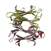
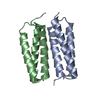
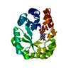
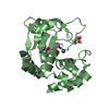
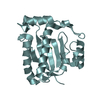
Structure visualization

Structure viewerMolecule:
MolmilJmol/JSmol

Downloads & links

-
Assembly

Deposited unit
A: Rhino
B: Rhino
C: GD21652
D: GD21652


Theoretical massNumber of molelcules
Total (without water)29,1274
Polymers29,1274
Non-polymers00
Water1086
1


  • Idetical with deposited unit
  • defined by author&software
  • Evidence: gel filtration
TypeNameSymmetry operationNumber
identity operation1_555x,y,z1
Buried area4440 Å2
ΔGint-39 kcal/mol
Surface area9610 Å2
MethodPISA
Unit cell
Length a, b, c (Å)46.893, 46.893, 250.610
Angle α, β, γ (deg.)90.00, 90.00, 90.00
Int Tables number91
Space group name H-MP4122

-
Components

#1: Protein Rhino


Mass: 7569.646 Da / Num. of mol.: 2 / Fragment: UNP residues 440-497
Source method: isolated from a genetically manipulated source
Source: (gene. exp.) Drosophila simulans (fry) / Gene: rhi, Dsim\GD27086, Dsimw501_GD27086, GD25457 / Production host: Escherichia coli BL21(DE3) (bacteria) / Strain (production host): BL21(DE3) / References: UniProt: Q49BI5
#2: Protein GD21652


Mass: 6993.971 Da / Num. of mol.: 2 / Fragment: UNP residues 1-60
Source method: isolated from a genetically manipulated source
Details: In the electron density maps we only observed the main chain of 45Q, so we used Alanine instead.
Source: (gene. exp.) Drosophila simulans (fry) / Gene: Dsim\GD21652, Dsim_GD21652, GD21652 / Production host: Escherichia coli BL21(DE3) (bacteria) / Strain (production host): BL21(DE3) / References: UniProt: B4Q3Z0
#3: Water ChemComp-HOH / water


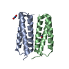
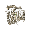
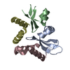
Mass: 18.015 Da / Num. of mol.: 6 / Source method: isolated from a natural source / Formula: H2O

-
Experimental details

-
Experiment

ExperimentMethod: X-RAY DIFFRACTION / Number of used crystals: 1

-
Sample preparation

CrystalDensity Matthews: 2.36 Å3/Da / Density % sol: 47.99 %
Crystal growTemperature: 290 K / Method: vapor diffusion, hanging drop / pH: 7 / Details: 2.5 M NaCl, 0.1 M Tris (pH7.0), 0.2 M MgCl2

-
Data collection

DiffractionMean temperature: 100 K
Diffraction sourceSource: SYNCHROTRON / Site: SSRF / Beamline: BL19U1 / Wavelength: 0.978 Å
DetectorType: DECTRIS PILATUS 6M / Detector: PIXEL / Date: Jun 10, 2017
RadiationProtocol: SINGLE WAVELENGTH / Monochromatic (M) / Laue (L): M / Scattering type: x-ray
Radiation wavelengthWavelength: 0.978 Å / Relative weight: 1
ReflectionResolution: 2.7→30 Å / Num. obs: 14516 / % possible obs: 99.8 % / Redundancy: 21.4 % / Net I/σ(I): 39
Reflection shellResolution: 2.7→2.8 Å / Redundancy: 12.9 % / Mean I/σ(I) obs: 3.6 / Num. unique obs: 14516 / % possible all: 98.5

-
Processing

Software
NameVersionClassification
PHENIX1.8.4_1496refinement
HKL-3000data reduction
HKL-3000data scaling
PHENIXphasing
RefinementMethod to determine structure: SAD / Resolution: 2.705→23.942 Å / SU ML: 0.42 / Cross valid method: FREE R-VALUE / σ(F): 0 / Phase error: 34.01
RfactorNum. reflection% reflection
Rfree0.3111 1465 10.09 %
Rwork0.2426 --
obs0.2494 14516 99.38 %
Solvent computationShrinkage radii: 0.9 Å / VDW probe radii: 1.11 Å
Refinement stepCycle: LAST / Resolution: 2.705→23.942 Å
ProteinNucleic acidLigandSolventTotal
Num. atoms1439 0 0 6 1445
Refine LS restraints
Refine-IDTypeDev idealNumber
X-RAY DIFFRACTIONf_bond_d0.011475
X-RAY DIFFRACTIONf_angle_d1.3391984
X-RAY DIFFRACTIONf_dihedral_angle_d15.453530
X-RAY DIFFRACTIONf_chiral_restr0.054211
X-RAY DIFFRACTIONf_plane_restr0.006240
LS refinement shell
Resolution (Å)Rfactor RfreeNum. reflection RfreeRfactor RworkNum. reflection RworkRefine-ID% reflection obs (%)
2.7047-2.80120.37371470.33091252X-RAY DIFFRACTION95
2.8012-2.91320.33211490.29851327X-RAY DIFFRACTION100
2.9132-3.04550.3731440.28411278X-RAY DIFFRACTION100
3.0455-3.20570.31821480.27091312X-RAY DIFFRACTION100
3.2057-3.4060.38391470.26371317X-RAY DIFFRACTION100
3.406-3.66820.31521500.25041318X-RAY DIFFRACTION100
3.6682-4.03570.27731420.23131314X-RAY DIFFRACTION100
4.0357-4.61610.3031460.20261315X-RAY DIFFRACTION100
4.6161-5.80210.24721430.22141317X-RAY DIFFRACTION100
5.8021-23.94340.341490.25261301X-RAY DIFFRACTION99
Refinement TLS params.Method: refined / Origin x: 23.0879 Å / Origin y: -6.7718 Å / Origin z: -19.5351 Å
111213212223313233
T0.4928 Å2-0.2042 Å2-0.0432 Å2-0.5815 Å2-0.001 Å2--0.4515 Å2
L1.0463 °2-0.576 °2-0.7101 °2-1.7877 °20.0794 °2--1.6218 °2
S0.0325 Å °-0.291 Å °0.1728 Å °-0.1174 Å °0.2702 Å °0.1536 Å °0.0642 Å °0.0837 Å °0.0032 Å °
Refinement TLS groupSelection details: all

+
About Yorodumi

-
News

-
Feb 9, 2022. New format data for meta-information of EMDB entries

New format data for meta-information of EMDB entries

  • Version 3 of the EMDB header file is now the official format.
  • The previous official version 1.9 will be removed from the archive.

Related info.:EMDB header

External links:wwPDB to switch to version 3 of the EMDB data model

-
Aug 12, 2020. Covid-19 info

Covid-19 info

URL: https://pdbj.org/emnavi/covid19.php

New page: Covid-19 featured information page in EM Navigator.

Related info.:Covid-19 info / Mar 5, 2020. Novel coronavirus structure data

+
Mar 5, 2020. Novel coronavirus structure data

Novel coronavirus structure data

Related info.:Yorodumi Speices / Aug 12, 2020. Covid-19 info

External links:COVID-19 featured content - PDBj / Molecule of the Month (242):Coronavirus Proteases

+
Jan 31, 2019. EMDB accession codes are about to change! (news from PDBe EMDB page)

EMDB accession codes are about to change! (news from PDBe EMDB page)

  • The allocation of 4 digits for EMDB accession codes will soon come to an end. Whilst these codes will remain in use, new EMDB accession codes will include an additional digit and will expand incrementally as the available range of codes is exhausted. The current 4-digit format prefixed with “EMD-” (i.e. EMD-XXXX) will advance to a 5-digit format (i.e. EMD-XXXXX), and so on. It is currently estimated that the 4-digit codes will be depleted around Spring 2019, at which point the 5-digit format will come into force.
  • The EM Navigator/Yorodumi systems omit the EMD- prefix.

Related info.:Q: What is EMD? / ID/Accession-code notation in Yorodumi/EM Navigator

External links:EMDB Accession Codes are Changing Soon! / Contact to PDBj

+
Jul 12, 2017. Major update of PDB

Major update of PDB

  • wwPDB released updated PDB data conforming to the new PDBx/mmCIF dictionary.
  • This is a major update changing the version number from 4 to 5, and with Remediation, in which all the entries are updated.
  • In this update, many items about electron microscopy experimental information are reorganized (e.g. em_software).
  • Now, EM Navigator and Yorodumi are based on the updated data.

External links:wwPDB Remediation / Enriched Model Files Conforming to OneDep Data Standards Now Available in the PDB FTP Archive

-
Yorodumi

Thousand views of thousand structures

  • Yorodumi is a browser for structure data from EMDB, PDB, SASBDB, etc.
  • This page is also the successor to EM Navigator detail page, and also detail information page/front-end page for Omokage search.
  • The word "yorodu" (or yorozu) is an old Japanese word meaning "ten thousand". "mi" (miru) is to see.

Related info.:EMDB / PDB / SASBDB / Comparison of 3 databanks / Yorodumi Search / Aug 31, 2016. New EM Navigator & Yorodumi / Yorodumi Papers / Jmol/JSmol / Function and homology information / Changes in new EM Navigator and Yorodumi

Read more