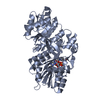
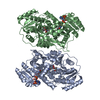
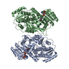
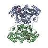
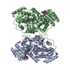
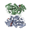
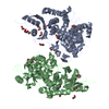
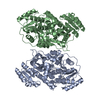
[English] 日本語
Yorodumi
- PDB-5xww: Substrate-bound Structure of G355T/Q364H mutant of a Ketoreductas... -

+
Open data


ID or keywords:

Loading...

-
Basic information

Entry
Database: PDB / ID: 5xww
TitleSubstrate-bound Structure of G355T/Q364H mutant of a Ketoreductase from amphotericin Polyketide Synthases
ComponentsAmphB
KeywordsOXIDOREDUCTASE / modular polyketide synthease / ketoreductase / mutant
Function / homology
Function and homology information


macrolide biosynthetic process / phosphopantetheine binding / 3-oxoacyl-[acyl-carrier-protein] synthase activity / fatty acid biosynthetic process / nucleotide binding / identical protein binding
Similarity search - Function
Polyketide synthase dimerisation element domain / Polyketide synthase dimerisation element domain / Polyketide synthase, docking domain / Erythronolide synthase docking domain / PKS_PP_betabranch / Polyketide synthase, ketoreductase domain / KR domain / Polyketide synthase, C-terminal extension / Ketoacyl-synthetase C-terminal extension / Malonyl-CoA ACP transacylase, ACP-binding ...Polyketide synthase dimerisation element domain / Polyketide synthase dimerisation element domain / Polyketide synthase, docking domain / Erythronolide synthase docking domain / PKS_PP_betabranch / Polyketide synthase, ketoreductase domain / KR domain / Polyketide synthase, C-terminal extension / Ketoacyl-synthetase C-terminal extension / Malonyl-CoA ACP transacylase, ACP-binding / PKS_KR / Acyl transferase domain superfamily / Acyl transferase / Acyl transferase domain / Acyl transferase domain in polyketide synthase (PKS) enzymes. / Acyl transferase/acyl hydrolase/lysophospholipase / Ketosynthase family 3 (KS3) domain profile. / Polyketide synthase, phosphopantetheine-binding domain / Phosphopantetheine attachment site / Beta-ketoacyl synthase / Beta-ketoacyl synthase, active site / Ketosynthase family 3 (KS3) active site signature. / Polyketide synthase, beta-ketoacyl synthase domain / Beta-ketoacyl synthase, N-terminal / Beta-ketoacyl synthase, C-terminal / Beta-ketoacyl synthase, N-terminal domain / Beta-ketoacyl synthase, C-terminal domain / Phosphopantetheine attachment site / Thiolase-like / Phosphopantetheine attachment site. / Phosphopantetheine attachment site / ACP-like superfamily / Carrier protein (CP) domain profile. / Phosphopantetheine binding ACP domain / NAD(P)-binding Rossmann-like Domain / NAD(P)-binding domain superfamily / Rossmann fold / 3-Layer(aba) Sandwich / Alpha Beta
Similarity search - Domain/homology
Chem-8H6 / NADP NICOTINAMIDE-ADENINE-DINUCLEOTIDE PHOSPHATE / AmphB
Similarity search - Component
Biological speciesStreptomyces nodosus (bacteria)
MethodX-RAY DIFFRACTION / SYNCHROTRON / MOLECULAR REPLACEMENT / Resolution: 1.96 Å
AuthorsLiu, C. / Zheng, J.
Funding support China, 3items
OrganizationGrant numberCountry
National Program on Key Basic Research Project2013CB734002 China
National Natural Science Foundation of China31370101 China
National Natural Science Foundation of China31570056 China
CitationJournal: J. Struct. Biol. / Year: 2018
Title: Substrate-bound structures of a ketoreductase from amphotericin modular polyketide synthase.
Authors: Liu, C. / Yuan, M. / Xu, X. / Wang, L. / Keatinge-Clay, A.T. / Deng, Z. / Lin, S. / Zheng, J.
History
DepositionJun 30, 2017Deposition site: PDBJ / Processing site: PDBJ
Revision 1.0Jun 6, 2018Provider: repository / Type: Initial release
Revision 1.1Jul 4, 2018Group: Data collection / Database references / Category: citation
Item: _citation.journal_volume / _citation.page_first / _citation.page_last
Revision 1.2Nov 22, 2023Group: Data collection / Database references / Refinement description
Category: chem_comp_atom / chem_comp_bond ...chem_comp_atom / chem_comp_bond / database_2 / pdbx_initial_refinement_model / refine_hist / struct_ncs_dom_lim
Item: _database_2.pdbx_DOI / _database_2.pdbx_database_accession ..._database_2.pdbx_DOI / _database_2.pdbx_database_accession / _refine_hist.d_res_low / _struct_ncs_dom_lim.beg_auth_comp_id / _struct_ncs_dom_lim.beg_label_asym_id / _struct_ncs_dom_lim.beg_label_comp_id / _struct_ncs_dom_lim.beg_label_seq_id / _struct_ncs_dom_lim.end_auth_comp_id / _struct_ncs_dom_lim.end_label_asym_id / _struct_ncs_dom_lim.end_label_comp_id / _struct_ncs_dom_lim.end_label_seq_id

-
Structure visualization

Structure viewerMolecule:
MolmilJmol/JSmol

Downloads & links

-
Assembly

Deposited unit
A: AmphB
B: AmphB
hetero molecules


Theoretical massNumber of molelcules
Total (without water)105,2256
Polymers102,9582
Non-polymers2,2684
Water1,838102
1


  • Idetical with deposited unit
  • defined by author
  • Evidence: gel filtration
TypeNameSymmetry operationNumber
identity operation1_555x,y,z1
Buried area3650 Å2
ΔGint-12 kcal/mol
Surface area34480 Å2
Unit cell
Length a, b, c (Å)61.066, 63.627, 71.586
Angle α, β, γ (deg.)72.67, 67.21, 89.88
Int Tables number1
Space group name H-MP1
Noncrystallographic symmetry (NCS)NCS domain:
IDEns-IDDetails
11A
21B

NCS domain segments:

Component-ID: 0 / Ens-ID: 1 / Beg auth comp-ID: GLY / Beg label comp-ID: GLY / End auth comp-ID: SER / End label comp-ID: SER / Refine code: 0 / Auth seq-ID: -3 - 473 / Label seq-ID: 18 - 494

Dom-IDAuth asym-IDLabel asym-ID
1AA
2BB

-
Components

#1: Protein AmphB


Mass: 51478.766 Da / Num. of mol.: 2 / Fragment: UNP RESIDUES 2529-3003 / Mutation: G355T, Q364H
Source method: isolated from a genetically manipulated source
Source: (gene. exp.) Streptomyces nodosus (bacteria) / Gene: amphB / Production host: Escherichia coli (E. coli) / References: UniProt: Q93NW7
#2: Chemical ChemComp-NAP / NADP NICOTINAMIDE-ADENINE-DINUCLEOTIDE PHOSPHATE / 2'-MONOPHOSPHOADENOSINE 5'-DIPHOSPHORIBOSE / Nicotinamide adenine dinucleotide phosphate


Mass: 743.405 Da / Num. of mol.: 2 / Source method: obtained synthetically / Formula: C21H28N7O17P3
#3: Chemical ChemComp-8H6 / S-[2-[3-[[(2R)-3,3-dimethyl-2,4-bis(oxidanyl)butanoyl]amino]propanoylamino]ethyl] (2R)-2-methyl-3-oxidanylidene-pentanethioate


Mass: 390.495 Da / Num. of mol.: 2 / Source method: obtained synthetically / Formula: C17H30N2O6S / Feature type: SUBJECT OF INVESTIGATION
#4: Water ChemComp-HOH / water / Water


Mass: 18.015 Da / Num. of mol.: 102 / Source method: isolated from a natural source / Formula: H2O

-
Experimental details

-
Experiment

ExperimentMethod: X-RAY DIFFRACTION / Number of used crystals: 1

-
Sample preparation

CrystalDensity Matthews: 2.36 Å3/Da / Density % sol: 48 %
Crystal growTemperature: 293 K / Method: vapor diffusion / pH: 7.5 / Details: 3.4 M ammonium sulfate, 0.1 M HEPES, pH 7.5

-
Data collection

DiffractionMean temperature: 100 K
Diffraction sourceSource: SYNCHROTRON / Site: SSRF / Beamline: BL18U1 / Wavelength: 0.9778 Å
DetectorType: DECTRIS PILATUS3 6M / Detector: PIXEL / Date: Dec 21, 2016
RadiationProtocol: SINGLE WAVELENGTH / Monochromatic (M) / Laue (L): M / Scattering type: x-ray
Radiation wavelengthWavelength: 0.9778 Å / Relative weight: 1
ReflectionResolution: 1.96→62.48 Å / Num. obs: 63286 / % possible obs: 94.6 % / Redundancy: 1.8 % / Rmerge(I) obs: 0.1 / Net I/σ(I): 8.6
Reflection shellResolution: 1.96→1.99 Å / Redundancy: 1.6 % / Rmerge(I) obs: 0.368 / Mean I/σ(I) obs: 2 / Num. unique obs: 3033 / % possible all: 90.6

-
Processing

Software
NameVersionClassification
REFMAC5.8.0151refinement
HKL-2000data reduction
HKL-2000data scaling
PHASERphasing
RefinementMethod to determine structure: MOLECULAR REPLACEMENT
Starting model: 3MJE
Resolution: 1.96→50 Å / Cor.coef. Fo:Fc: 0.957 / Cor.coef. Fo:Fc free: 0.939 / SU B: 8.716 / SU ML: 0.116 / Cross valid method: THROUGHOUT / ESU R: 0.176 / ESU R Free: 0.153 / Stereochemistry target values: MAXIMUM LIKELIHOOD / Details: HYDROGENS HAVE BEEN ADDED IN THE RIDING POSITIONS
RfactorNum. reflection% reflectionSelection details
Rfree0.21656 3200 5.1 %RANDOM
Rwork0.1803 ---
obs0.18214 60085 94.15 %-
Solvent computationIon probe radii: 0.8 Å / Shrinkage radii: 0.8 Å / VDW probe radii: 1.2 Å / Solvent model: MASK
Displacement parametersBiso mean: 31.437 Å2
Baniso -1Baniso -2Baniso -3
1-0.87 Å20 Å20.11 Å2
2---0.55 Å2-0.9 Å2
3----0.51 Å2
Refinement stepCycle: 1 / Resolution: 1.96→50 Å
ProteinNucleic acidLigandSolventTotal
Num. atoms6946 0 148 102 7196
Refine LS restraints
Refine-IDTypeDev idealDev ideal targetNumber
X-RAY DIFFRACTIONr_bond_refined_d0.0070.0197242
X-RAY DIFFRACTIONr_bond_other_d00.026892
X-RAY DIFFRACTIONr_angle_refined_deg1.0881.9839910
X-RAY DIFFRACTIONr_angle_other_deg0.58315762
X-RAY DIFFRACTIONr_dihedral_angle_1_deg6.7315952
X-RAY DIFFRACTIONr_dihedral_angle_2_deg34.09522.867286
X-RAY DIFFRACTIONr_dihedral_angle_3_deg14.05151018
X-RAY DIFFRACTIONr_dihedral_angle_4_deg17.5161564
X-RAY DIFFRACTIONr_chiral_restr0.0620.21144
X-RAY DIFFRACTIONr_gen_planes_refined0.0160.0218328
X-RAY DIFFRACTIONr_gen_planes_other0.0030.021604
X-RAY DIFFRACTIONr_nbd_refined
X-RAY DIFFRACTIONr_nbd_other
X-RAY DIFFRACTIONr_nbtor_refined
X-RAY DIFFRACTIONr_nbtor_other
X-RAY DIFFRACTIONr_xyhbond_nbd_refined
X-RAY DIFFRACTIONr_xyhbond_nbd_other
X-RAY DIFFRACTIONr_metal_ion_refined
X-RAY DIFFRACTIONr_metal_ion_other
X-RAY DIFFRACTIONr_symmetry_vdw_refined
X-RAY DIFFRACTIONr_symmetry_vdw_other
X-RAY DIFFRACTIONr_symmetry_hbond_refined
X-RAY DIFFRACTIONr_symmetry_hbond_other
X-RAY DIFFRACTIONr_symmetry_metal_ion_refined
X-RAY DIFFRACTIONr_symmetry_metal_ion_other
X-RAY DIFFRACTIONr_mcbond_it2.7912.6753814
X-RAY DIFFRACTIONr_mcbond_other2.7912.6743813
X-RAY DIFFRACTIONr_mcangle_it3.7893.9924764
X-RAY DIFFRACTIONr_mcangle_other3.7893.9934765
X-RAY DIFFRACTIONr_scbond_it4.1043.2063428
X-RAY DIFFRACTIONr_scbond_other4.1043.2063429
X-RAY DIFFRACTIONr_scangle_it
X-RAY DIFFRACTIONr_scangle_other6.2184.6055147
X-RAY DIFFRACTIONr_long_range_B_refined8.2152.07830811
X-RAY DIFFRACTIONr_long_range_B_other8.21352.08730766
X-RAY DIFFRACTIONr_rigid_bond_restr
X-RAY DIFFRACTIONr_sphericity_free
X-RAY DIFFRACTIONr_sphericity_bonded
Refine LS restraints NCS

Ens-ID: 1 / Number: 28478 / Refine-ID: X-RAY DIFFRACTION / Type: interatomic distance / Rms dev position: 0.1 Å / Weight position: 0.05

Dom-IDAuth asym-ID
1A
2B
LS refinement shellResolution: 1.963→2.014 Å / Total num. of bins used: 20
RfactorNum. reflection% reflection
Rfree0.266 202 -
Rwork0.227 4089 -
obs--86.23 %
Refinement TLS params.

Method: refined / Refine-ID: X-RAY DIFFRACTION

IDL112)L122)L132)L222)L232)L332)S11 (Å °)S12 (Å °)S13 (Å °)S21 (Å °)S22 (Å °)S23 (Å °)S31 (Å °)S32 (Å °)S33 (Å °)T112)T122)T132)T222)T232)T332)Origin x (Å)Origin y (Å)Origin z (Å)
10.0924-0.0151-0.02170.26480.10530.10030.00210.0039-0.01250.01010.005-0.0177-0.0121-0.0007-0.00710.0193-0.0024-0.00370.01610.00470.0056-10.2922.0917-17.0972
20.07840.0057-0.00140.24370.08880.1155-0.0023-0.00610.008-0.01180.0012-0.01570.0045-0.00730.00110.02510.0024-0.00210.00790.00180.0051-13.0296-16.364223.6962
Refinement TLS group
IDRefine-IDRefine TLS-IDAuth asym-IDAuth seq-ID
1X-RAY DIFFRACTION1A-3 - 473
2X-RAY DIFFRACTION2B-3 - 473

+
About Yorodumi

-
News

-
Feb 9, 2022. New format data for meta-information of EMDB entries

New format data for meta-information of EMDB entries

  • Version 3 of the EMDB header file is now the official format.
  • The previous official version 1.9 will be removed from the archive.

Related info.:EMDB header

External links:wwPDB to switch to version 3 of the EMDB data model

-
Aug 12, 2020. Covid-19 info

Covid-19 info

URL: https://pdbj.org/emnavi/covid19.php

New page: Covid-19 featured information page in EM Navigator.

Related info.:Covid-19 info / Mar 5, 2020. Novel coronavirus structure data

+
Mar 5, 2020. Novel coronavirus structure data

Novel coronavirus structure data

Related info.:Yorodumi Speices / Aug 12, 2020. Covid-19 info

External links:COVID-19 featured content - PDBj / Molecule of the Month (242):Coronavirus Proteases

+
Jan 31, 2019. EMDB accession codes are about to change! (news from PDBe EMDB page)

EMDB accession codes are about to change! (news from PDBe EMDB page)

  • The allocation of 4 digits for EMDB accession codes will soon come to an end. Whilst these codes will remain in use, new EMDB accession codes will include an additional digit and will expand incrementally as the available range of codes is exhausted. The current 4-digit format prefixed with “EMD-” (i.e. EMD-XXXX) will advance to a 5-digit format (i.e. EMD-XXXXX), and so on. It is currently estimated that the 4-digit codes will be depleted around Spring 2019, at which point the 5-digit format will come into force.
  • The EM Navigator/Yorodumi systems omit the EMD- prefix.

Related info.:Q: What is EMD? / ID/Accession-code notation in Yorodumi/EM Navigator

External links:EMDB Accession Codes are Changing Soon! / Contact to PDBj

+
Jul 12, 2017. Major update of PDB

Major update of PDB

  • wwPDB released updated PDB data conforming to the new PDBx/mmCIF dictionary.
  • This is a major update changing the version number from 4 to 5, and with Remediation, in which all the entries are updated.
  • In this update, many items about electron microscopy experimental information are reorganized (e.g. em_software).
  • Now, EM Navigator and Yorodumi are based on the updated data.

External links:wwPDB Remediation / Enriched Model Files Conforming to OneDep Data Standards Now Available in the PDB FTP Archive

-
Yorodumi

Thousand views of thousand structures

  • Yorodumi is a browser for structure data from EMDB, PDB, SASBDB, etc.
  • This page is also the successor to EM Navigator detail page, and also detail information page/front-end page for Omokage search.
  • The word "yorodu" (or yorozu) is an old Japanese word meaning "ten thousand". "mi" (miru) is to see.

Related info.:EMDB / PDB / SASBDB / Comparison of 3 databanks / Yorodumi Search / Aug 31, 2016. New EM Navigator & Yorodumi / Yorodumi Papers / Jmol/JSmol / Function and homology information / Changes in new EM Navigator and Yorodumi

Read more