 Movie
Movie Controller


+ Open data

- Basic information
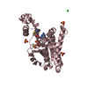
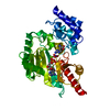
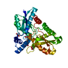
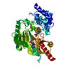
| Entry | Database: PDB / ID: 5xtu | ||||||
|---|---|---|---|---|---|---|---|
| Title | Crystal Structure of GDSL Esterase of Photobacterium sp. J15 | ||||||
 Components Components | GDSL-family esterase | ||||||
 Keywords Keywords | HYDROLASE / GDSL / SGNH / esterase / Photobacterium | ||||||
| Function / homology | GDSL lipase/esterase / GDSL-like Lipase/Acylhydrolase / SGNH hydrolase superfamily / hydrolase activity, acting on ester bonds / CACODYLATE ION / DI(HYDROXYETHYL)ETHER / PHOSPHATE ION / GDSL-family esterase Function and homology information Function and homology information | ||||||
| Biological species |  Photobacterium sp. J15 (bacteria) Photobacterium sp. J15 (bacteria) | ||||||
| Method |  X-RAY DIFFRACTION / SAD / Resolution: 1.38 Å X-RAY DIFFRACTION / SAD / Resolution: 1.38 Å | ||||||
 Authors Authors | Mazlan, S.N.H.S. / Jonet, M.A. / Leow, T.C. / Ali, M.S.M. / Rahman, R.N.Z.R.A. | ||||||
 Citation Citation | Journal: Int. J. Biol. Macromol. / Year: 2018 Title: Crystallization and structure elucidation of GDSL esterase of Photobacterium sp. J15. Authors: Mazlan, S.N.H.S. / Ali, M.S.M. / Rahman, R.N.Z.R.A. / Sabri, S. / Jonet, M.A. / Leow, T.C. | ||||||
| History |
|
- Structure visualization
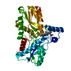
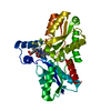
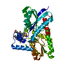
| Structure viewer | Molecule: MolmilJmol/JSmol |
|---|
- Downloads & links
-Download
| PDBx/mmCIF format | 5xtu.cif.gz | 92.6 KB | Display | PDBx/mmCIF format |
|---|---|---|---|---|
| PDB format | pdb5xtu.ent.gz | 66.8 KB | Display | PDB format |
| PDBx/mmJSON format | 5xtu.json.gz | Tree view | PDBx/mmJSON format | |
| Others |  Other downloads Other downloads |
-Validation report
| Arichive directory | https://data.pdbj.org/pub/pdb/validation_reports/xt/5xtuftp://data.pdbj.org/pub/pdb/validation_reports/xt/5xtu | HTTPS FTP |
|---|
-Related structure data
| Similar structure data |
|---|


-Links
 PDBj
PDBj


- Assembly
| Deposited unit | 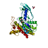
| ||||||||
|---|---|---|---|---|---|---|---|---|---|
| 1 |
| ||||||||
| Unit cell |
|
-Components
-Protein , 1 types, 1 molecules A
| #1: Protein | Mass: 39101.695 Da / Num. of mol.: 1 Source method: isolated from a genetically manipulated source Source: (gene. exp.) Photobacterium sp. J15(2011) (bacteria)Strain: 2011 / Plasmid: pET32b / Production host: |
|---|
-Non-polymers , 7 types, 444 molecules 


| #2: Chemical | ChemComp-PEG /  Mass: 106.120 Da / Num. of mol.: 1 / Source method: obtained synthetically / Formula: C4H10O3 / Feature type: SUBJECT OF INVESTIGATION Mass: 106.120 Da / Num. of mol.: 1 / Source method: obtained synthetically / Formula: C4H10O3 / Feature type: SUBJECT OF INVESTIGATION | ||||||||
|---|---|---|---|---|---|---|---|---|---|
| #3: Chemical | ChemComp-CL /  Mass: 35.453 Da / Num. of mol.: 1 / Source method: obtained synthetically / Formula: Cl / Feature type: SUBJECT OF INVESTIGATION Mass: 35.453 Da / Num. of mol.: 1 / Source method: obtained synthetically / Formula: Cl / Feature type: SUBJECT OF INVESTIGATION | ||||||||
| #4: Chemical |  Mass: 136.989 Da / Num. of mol.: 2 / Source method: obtained synthetically / Formula: C2H6AsO2 / Feature type: SUBJECT OF INVESTIGATION Mass: 136.989 Da / Num. of mol.: 2 / Source method: obtained synthetically / Formula: C2H6AsO2 / Feature type: SUBJECT OF INVESTIGATION#5: Chemical | ChemComp-CA / |  Mass: 40.078 Da / Num. of mol.: 1 / Source method: obtained synthetically / Formula: Ca / Feature type: SUBJECT OF INVESTIGATION Mass: 40.078 Da / Num. of mol.: 1 / Source method: obtained synthetically / Formula: Ca / Feature type: SUBJECT OF INVESTIGATION#6: Chemical | ChemComp-EDO / |  Mass: 62.068 Da / Num. of mol.: 1 / Source method: obtained synthetically / Formula: C2H6O2 / Feature type: SUBJECT OF INVESTIGATION Mass: 62.068 Da / Num. of mol.: 1 / Source method: obtained synthetically / Formula: C2H6O2 / Feature type: SUBJECT OF INVESTIGATION#7: Chemical |  Mass: 94.971 Da / Num. of mol.: 2 / Source method: obtained synthetically / Formula: PO4 / Feature type: SUBJECT OF INVESTIGATION Mass: 94.971 Da / Num. of mol.: 2 / Source method: obtained synthetically / Formula: PO4 / Feature type: SUBJECT OF INVESTIGATION#8: Water | ChemComp-HOH / | Mass: 18.015 Da / Num. of mol.: 436 / Source method: isolated from a natural source / Formula: H2O |
-Details
| Has protein modification | N |
|---|
-Experimental details
-Experiment
| Experiment | Method: X-RAY DIFFRACTION / Number of used crystals: 1 |
|---|
- Sample preparation
| Crystal | Density Matthews: 2.42 Å3/Da / Density % sol: 44.2 % |
|---|---|
| Crystal grow | Temperature: 293 K / Method: vapor diffusion, sitting drop / pH: 6.5 Details: 0.10 M ammonium sulphate, 0.15 M sodium cacodylate trihydrate pH 6.5, and 20% PEG 8000 |
-Data collection
| Diffraction | Mean temperature: 100 K |
|---|---|
| Diffraction source | Source: ROTATING ANODE / Type: RIGAKU / Wavelength: 1.54 Å |
| Detector | Type: RIGAKU / Detector: CCD / Date: Jul 22, 2016 |
| Radiation | Protocol: SINGLE WAVELENGTH / Monochromatic (M) / Laue (L): M / Scattering type: x-ray |
| Radiation wavelength | Wavelength: 1.54 Å / Relative weight: 1 |
| Reflection | Resolution: 1.38→56.23 Å / Num. obs: 70491 / % possible obs: 98.2 % / Redundancy: 2 % / CC1/2: 0.912 / Rmerge(I) obs: 0.07 / Rpim(I) all: 0.07 / Net I/σ(I): 25 |
| Reflection shell | Resolution: 1.38→1.43 Å / Redundancy: 1.98 % / Rmerge(I) obs: 0.193 / Mean I/σ(I) obs: 5.7 / % possible all: 95.3 |
- Processing
| Software |
| ||||||||||||||||||||||||||||||||||||||||||||||||||||||||||||
|---|---|---|---|---|---|---|---|---|---|---|---|---|---|---|---|---|---|---|---|---|---|---|---|---|---|---|---|---|---|---|---|---|---|---|---|---|---|---|---|---|---|---|---|---|---|---|---|---|---|---|---|---|---|---|---|---|---|---|---|---|---|
| Refinement | Method to determine structure: SAD / Resolution: 1.38→56.23 Å / Cor.coef. Fo:Fc: 0.966 / Cor.coef. Fo:Fc free: 0.958 / SU B: 0.773 / SU ML: 0.032 / Cross valid method: THROUGHOUT / σ(F): 0 / ESU R: 0.053 / ESU R Free: 0.054 Details: HYDROGENS HAVE BEEN ADDED IN THE RIDING POSITIONS U VALUES : REFINED INDIVIDUALLY
| ||||||||||||||||||||||||||||||||||||||||||||||||||||||||||||
| Solvent computation | Ion probe radii: 0.8 Å / Shrinkage radii: 0.8 Å / VDW probe radii: 1.2 Å | ||||||||||||||||||||||||||||||||||||||||||||||||||||||||||||
| Displacement parameters | Biso max: 66.98 Å2 / Biso mean: 12.254 Å2 / Biso min: 3.74 Å2
| ||||||||||||||||||||||||||||||||||||||||||||||||||||||||||||
| Refinement step | Cycle: final / Resolution: 1.38→56.23 Å
| ||||||||||||||||||||||||||||||||||||||||||||||||||||||||||||
| Refine LS restraints |
| ||||||||||||||||||||||||||||||||||||||||||||||||||||||||||||
| LS refinement shell | Resolution: 1.38→1.416 Å / Rfactor Rfree error: 0 / Total num. of bins used: 20
|
