- PDB-5xmy: Crystal structure of TAF3 PHD finger bound to H3K4me3Q5ser -
+
Open data
ID or keywords:
Loading...
-
Basic information
Entry
Database: PDB / ID: 5xmy
Title
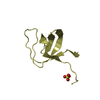
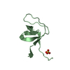
Crystal structure of TAF3 PHD finger bound to H3K4me3Q5ser
Components
Histone peptide H3(1-15)K4me3Q5ser
Transcription initiation factor TFIID subunit 3
Keywords
TRANSCRIPTION / zinc finger
Function / homology
Function and homology information
maintenance of protein location in nucleus / histone H3K4me3 reader activity / transcription factor TFIID complex / RNA polymerase II general transcription initiation factor activity / HIV Transcription Initiation / RNA Polymerase II HIV Promoter Escape / Transcription of the HIV genome / RNA Polymerase II Promoter Escape / RNA Polymerase II Transcription Pre-Initiation And Promoter Opening / RNA Polymerase II Transcription Initiation ...maintenance of protein location in nucleus / histone H3K4me3 reader activity / transcription factor TFIID complex / RNA polymerase II general transcription initiation factor activity / HIV Transcription Initiation / RNA Polymerase II HIV Promoter Escape / Transcription of the HIV genome / RNA Polymerase II Promoter Escape / RNA Polymerase II Transcription Pre-Initiation And Promoter Opening / RNA Polymerase II Transcription Initiation / RNA Polymerase II Transcription Initiation And Promoter Clearance / negative regulation of signal transduction by p53 class mediator / positive regulation of transcription initiation by RNA polymerase II / RNA polymerase II preinitiation complex assembly / transcription regulator inhibitor activity / RNA Polymerase II Pre-transcription Events / male germ cell nucleus / mRNA transcription by RNA polymerase II / p53 binding / nuclear membrane / Regulation of TP53 Activity through Phosphorylation / protein heterodimerization activity / negative regulation of transcription by RNA polymerase II / positive regulation of transcription by RNA polymerase II / zinc ion binding / nucleoplasm / nucleus Similarity search - Function
In the structure databanks used in Yorodumi, some data are registered as the other names, "COVID-19 virus" and "2019-nCoV". Here are the details of the virus and the list of structure data.
Jan 31, 2019. EMDB accession codes are about to change! (news from PDBe EMDB page)
EMDB accession codes are about to change! (news from PDBe EMDB page)
The allocation of 4 digits for EMDB accession codes will soon come to an end. Whilst these codes will remain in use, new EMDB accession codes will include an additional digit and will expand incrementally as the available range of codes is exhausted. The current 4-digit format prefixed with “EMD-” (i.e. EMD-XXXX) will advance to a 5-digit format (i.e. EMD-XXXXX), and so on. It is currently estimated that the 4-digit codes will be depleted around Spring 2019, at which point the 5-digit format will come into force.
The EM Navigator/Yorodumi systems omit the EMD- prefix.
Related info.:Q: What is EMD? / ID/Accession-code notation in Yorodumi/EM Navigator
Yorodumi is a browser for structure data from EMDB, PDB, SASBDB, etc.
This page is also the successor to EM Navigator detail page, and also detail information page/front-end page for Omokage search.
The word "yorodu" (or yorozu) is an old Japanese word meaning "ten thousand". "mi" (miru) is to see.
Related info.:EMDB / PDB / SASBDB / Comparison of 3 databanks / Yorodumi Search / Aug 31, 2016. New EM Navigator & Yorodumi / Yorodumi Papers / Jmol/JSmol / Function and homology information / Changes in new EM Navigator and Yorodumi
 Movie
Movie Controller


 Open data
 Basic information
 Components
 Keywords
 Function and homology information
 Homo sapiens (human)
 X-RAY DIFFRACTION /
  Authors
 Citation
 Structure visualization
 Downloads & links
 Other downloads


 PDBj
PDBj


 Assembly

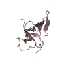


 Mass: 65.409 Da / Num. of mol.: 4 / Source method: obtained synthetically / Formula: Zn
 Mass: 18.015 Da / Num. of mol.: 107 / Source method: isolated from a natural source / Formula: H2O
 Sample preparation
 / Beamline: BL17U1 / Wavelength: 0.9792 Å
 Processing