 Movie
Movie Controller

[English] 日本語
 Yorodumi
- PDB-5xe8: Crystal Structure of the Peptidase Domain of Streptococcus mutans ComA -
+ Open data

- Basic information
| Entry | Database: PDB / ID: 5xe8 | ||||||
|---|---|---|---|---|---|---|---|
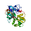
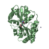
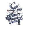
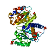
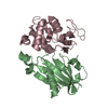
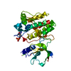
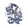
| Title | Crystal Structure of the Peptidase Domain of Streptococcus mutans ComA | ||||||
 Components Components | Putative ABC transporter, ATP-binding protein ComA | ||||||
 Keywords Keywords | HYDROLASE/HYDROLASE INHIBITOR / Peptidase / Quorum-sensing / HYDROLASE-HYDROLASE INHIBITOR complex | ||||||
| Function / homology |  Function and homology information Function and homology informationsingle-species submerged biofilm formation / ABC-type bacteriocin transporter activity / ATPase-coupled lipid transmembrane transporter activity / cysteine-type peptidase activity / protein transport / ATP hydrolysis activity / proteolysis / ATP binding / metal ion binding / plasma membrane Similarity search - Function | ||||||
| Biological species |  Streptococcus mutans serotype c (bacteria) Streptococcus mutans serotype c (bacteria) | ||||||
| Method |  X-RAY DIFFRACTION / SYNCHROTRON / MOLECULAR REPLACEMENT / Resolution: 3.1 Å X-RAY DIFFRACTION / SYNCHROTRON / MOLECULAR REPLACEMENT / Resolution: 3.1 Å | ||||||
 Authors Authors | Ishii, S. / Fukui, K. / Yokoshima, S. / Kumagai, K. / Beniyama, Y. / Kodama, T. / Fukuyama, T. / Okabe, T. / Nagano, T. / Kojima, H. / Yano, T. | ||||||
 Citation Citation | Journal: Sci Rep / Year: 2017 Title: High-throughput Screening of Small Molecule Inhibitors of the Streptococcus Quorum-sensing Signal Pathway Authors: Ishii, S. / Fukui, K. / Yokoshima, S. / Kumagai, K. / Beniyama, Y. / Kodama, T. / Fukuyama, T. / Okabe, T. / Nagano, T. / Kojima, H. / Yano, T. | ||||||
| History |
|
- Structure visualization
| Structure viewer | Molecule: MolmilJmol/JSmol |
|---|
- Downloads & links
-Download
| PDBx/mmCIF format | 5xe8.cif.gz | 120.4 KB | Display | PDBx/mmCIF format |
|---|---|---|---|---|
| PDB format | pdb5xe8.ent.gz | 92.6 KB | Display | PDB format |
| PDBx/mmJSON format | 5xe8.json.gz | Tree view | PDBx/mmJSON format | |
| Others |  Other downloads Other downloads |
-Validation report
| Arichive directory | https://data.pdbj.org/pub/pdb/validation_reports/xe/5xe8ftp://data.pdbj.org/pub/pdb/validation_reports/xe/5xe8 | HTTPS FTP |
|---|
-Related structure data
| Related structure data | 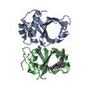 5xe9C  3k8uS C: citing same article ( S: Starting model for refinement |
|---|---|
| Similar structure data |


-Links
 PDBj
PDBj

- Assembly
| Deposited unit | 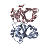
| ||||||||
|---|---|---|---|---|---|---|---|---|---|
| 1 |
| ||||||||
| Unit cell |
|
-Components
| #1: Protein | Mass: 17353.826 Da / Num. of mol.: 2 Source method: isolated from a genetically manipulated source Details: The first methionine and the last 16 residues are not identified in the crystal structure. Source: (gene. exp.) Streptococcus mutans serotype c (bacteria)Strain: ATCC 700610 / UA159 / Gene: SMU_286 / Plasmid: pET-21b / Production host: #2: Chemical |   Mass: 106.120 Da / Num. of mol.: 2 / Source method: obtained synthetically / Formula: C4H10O3 Mass: 106.120 Da / Num. of mol.: 2 / Source method: obtained synthetically / Formula: C4H10O3#3: Water | ChemComp-HOH / |  Mass: 18.015 Da / Num. of mol.: 1 / Source method: isolated from a natural source / Formula: H2O Mass: 18.015 Da / Num. of mol.: 1 / Source method: isolated from a natural source / Formula: H2O |
|---|
-Experimental details
-Experiment
| Experiment | Method: X-RAY DIFFRACTION / Number of used crystals: 1 |
|---|
- Sample preparation
| Crystal | Density Matthews: 2.45 Å3/Da / Density % sol: 49.84 % |
|---|---|
| Crystal grow | Temperature: 295 K / Method: vapor diffusion, sitting drop / pH: 5.5 Details: 100mM ammonium sulfate, 50mM bis-tris, 12.5%(W/v) polyethylene glycol 3,350 |
-Data collection
| Diffraction | Mean temperature: 100 K |
|---|---|
| Diffraction source | Source: SYNCHROTRON / Site: SPring-8  / Beamline: BL38B1 / Wavelength: 1 Å / Beamline: BL38B1 / Wavelength: 1 Å |
| Detector | Type: RAYONIX MX225HE / Detector: CCD / Date: Nov 9, 2015 |
| Radiation | Protocol: SINGLE WAVELENGTH / Monochromatic (M) / Laue (L): M / Scattering type: x-ray |
| Radiation wavelength | Wavelength: 1 Å / Relative weight: 1 |
| Reflection | Resolution: 2.8→50 Å / Num. obs: 6597 / % possible obs: 99.9 % / Redundancy: 6.8 % / Rmerge(I) obs: 0.069 / Net I/σ(I): 29 |
| Reflection shell | Resolution: 2.8→2.85 Å / Rmerge(I) obs: 0.557 |
- Processing
| Software |
| ||||||||||||||||||||||||||||||||||||||||
|---|---|---|---|---|---|---|---|---|---|---|---|---|---|---|---|---|---|---|---|---|---|---|---|---|---|---|---|---|---|---|---|---|---|---|---|---|---|---|---|---|---|
| Refinement | Method to determine structure: MOLECULAR REPLACEMENT Starting model: 3K8U Resolution: 3.1→31.551 Å / SU ML: 0.35 / Cross valid method: FREE R-VALUE / σ(F): 1.35 / Phase error: 28.31
| ||||||||||||||||||||||||||||||||||||||||
| Solvent computation | Shrinkage radii: 0.9 Å / VDW probe radii: 1.11 Å | ||||||||||||||||||||||||||||||||||||||||
| Refinement step | Cycle: LAST / Resolution: 3.1→31.551 Å
| ||||||||||||||||||||||||||||||||||||||||
| Refine LS restraints |
| ||||||||||||||||||||||||||||||||||||||||
| LS refinement shell |
| ||||||||||||||||||||||||||||||||||||||||
| Refinement TLS params. | Method: refined / Origin x: -3.0367 Å / Origin y: 2.5087 Å / Origin z: 42.7215 Å
| ||||||||||||||||||||||||||||||||||||||||
| Refinement TLS group | Selection details: all |
