 Movie
Movie Controller

[English] 日本語
 Yorodumi
- PDB-5x6q: Crystal structure of Pseudomonas fluorescens KMO in complex with ... -
+ Open data

- Basic information
| Entry | Database: PDB / ID: 5x6q | ||||||
|---|---|---|---|---|---|---|---|
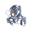
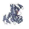
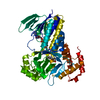
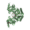
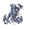
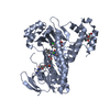
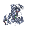
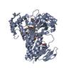
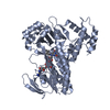
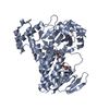
| Title | Crystal structure of Pseudomonas fluorescens KMO in complex with Ro 61-8048 | ||||||
 Components Components | Kynurenine 3-monooxygenase | ||||||
 Keywords Keywords | OXIDOREDUCTASE / monoxygenase | ||||||
| Function / homology |  Function and homology information Function and homology informationkynurenine 3-monooxygenase / kynurenine 3-monooxygenase activity / : / : / NAD(P)H oxidase H2O2-forming activity / L-tryptophan catabolic process / NAD+ biosynthetic process / quinolinate biosynthetic process / NAD+ metabolic process / FAD binding Similarity search - Function | ||||||
| Biological species |  Pseudomonas fluorescens (bacteria) Pseudomonas fluorescens (bacteria) | ||||||
| Method |  X-RAY DIFFRACTION / SYNCHROTRON / MOLECULAR REPLACEMENT / Resolution: 1.897 Å X-RAY DIFFRACTION / SYNCHROTRON / MOLECULAR REPLACEMENT / Resolution: 1.897 Å | ||||||
 Authors Authors | Kim, H.T. / Hwang, K.Y. | ||||||
 Citation Citation | Journal: Cell Chem Biol / Year: 2018 Title: Structural Basis for Inhibitor-Induced Hydrogen Peroxide Production by Kynurenine 3-Monooxygenase Authors: Kim, H.T. / Na, B.K. / Chung, J. / Kim, S. / Kwon, S.K. / Cha, H. / Son, J. / Cho, J.M. / Hwang, K.Y. | ||||||
| History |
|
- Structure visualization
| Structure viewer | Molecule: MolmilJmol/JSmol |
|---|
- Downloads & links
-Download
| PDBx/mmCIF format | 5x6q.cif.gz | 110.5 KB | Display | PDBx/mmCIF format |
|---|---|---|---|---|
| PDB format | pdb5x6q.ent.gz | 81.1 KB | Display | PDB format |
| PDBx/mmJSON format | 5x6q.json.gz | Tree view | PDBx/mmJSON format | |
| Others |  Other downloads Other downloads |
-Validation report
| Arichive directory | https://data.pdbj.org/pub/pdb/validation_reports/x6/5x6qftp://data.pdbj.org/pub/pdb/validation_reports/x6/5x6q | HTTPS FTP |
|---|
-Related structure data
| Related structure data |  5x68C  5x6pC  5x6rC  5fn0S C: citing same article ( S: Starting model for refinement |
|---|---|
| Similar structure data |
-Links
 PDBj
PDBj- Assembly
| Deposited unit | 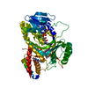
| ||||||||
|---|---|---|---|---|---|---|---|---|---|
| 1 |
| ||||||||
| Unit cell |
|
-Components
| #1: Protein | Mass: 50883.648 Da / Num. of mol.: 1 / Mutation: C252S,C461S Source method: isolated from a genetically manipulated source Source: (gene. exp.) Pseudomonas fluorescens (bacteria) / Gene: kmo, qbsGProduction host: References: UniProt: Q84HF5, kynurenine 3-monooxygenase | ||
|---|---|---|---|
| #2: Chemical | ChemComp-FAD /   Mass: 785.550 Da / Num. of mol.: 1 / Source method: obtained synthetically / Formula: C27H33N9O15P2 / Comment: FAD*YM Mass: 785.550 Da / Num. of mol.: 1 / Source method: obtained synthetically / Formula: C27H33N9O15P2 / Comment: FAD*YM | ||
| #3: Chemical |   Mass: 421.448 Da / Num. of mol.: 2 / Source method: obtained synthetically / Formula: C17H15N3O6S2 Mass: 421.448 Da / Num. of mol.: 2 / Source method: obtained synthetically / Formula: C17H15N3O6S2#4: Water | ChemComp-HOH / |  Mass: 18.015 Da / Num. of mol.: 203 / Source method: isolated from a natural source / Formula: H2O Mass: 18.015 Da / Num. of mol.: 203 / Source method: isolated from a natural source / Formula: H2O |
-Experimental details
-Experiment
| Experiment | Method: X-RAY DIFFRACTION / Number of used crystals: 1 |
|---|
- Sample preparation
| Crystal | Density Matthews: 2.28 Å3/Da / Density % sol: 45.97 % |
|---|---|
| Crystal grow | Temperature: 295 K / Method: vapor diffusion, hanging drop / pH: 6.5 Details: 0.08 M sodium cacodylate (pH 6.5), 0.16 M calcium acetate, 14.4% polyethylene glycol 8000, and 20% glycerol |
-Data collection
| Diffraction | Mean temperature: 100 K |
|---|---|
| Diffraction source | Source: SYNCHROTRON / Site: PAL/PLS  / Beamline: 5C (4A) / Wavelength: 1 Å / Beamline: 5C (4A) / Wavelength: 1 Å |
| Detector | Type: DECTRIS PILATUS3 S 6M / Detector: CCD / Date: May 1, 2016 |
| Radiation | Protocol: SINGLE WAVELENGTH / Monochromatic (M) / Laue (L): M / Scattering type: x-ray |
| Radiation wavelength | Wavelength: 1 Å / Relative weight: 1 |
| Reflection | Resolution: 1.897→33.653 Å / Num. obs: 35769 / % possible obs: 97.64 % / Redundancy: 3.2 % / Rmerge(I) obs: 0.06 / Rpim(I) all: 0.047 / Rrim(I) all: 0.084 / Rsym value: 0.069 / Χ2: 2.615 / Net I/σ(I): 30.96 |
| Reflection shell | Resolution: 1.9→1.93 Å / Redundancy: 3.4 % / Rmerge(I) obs: 0.452 / Mean I/σ(I) obs: 4.6 / Num. unique obs: 1786 / CC1/2: 0.838 / Rpim(I) all: 0.241 / Rrim(I) all: 0.447 / Rsym value: 0.375 / Χ2: 1.048 / % possible all: 99.9 |
- Processing
| Software |
| ||||||||||||||||||||||||||||||||||||||||||||||||||||||||||||||||||||||||||||||||||||||||||||||||||
|---|---|---|---|---|---|---|---|---|---|---|---|---|---|---|---|---|---|---|---|---|---|---|---|---|---|---|---|---|---|---|---|---|---|---|---|---|---|---|---|---|---|---|---|---|---|---|---|---|---|---|---|---|---|---|---|---|---|---|---|---|---|---|---|---|---|---|---|---|---|---|---|---|---|---|---|---|---|---|---|---|---|---|---|---|---|---|---|---|---|---|---|---|---|---|---|---|---|---|---|
| Refinement | Method to determine structure: MOLECULAR REPLACEMENT Starting model: 5FN0 Resolution: 1.897→33.653 Å / SU ML: 0.22 / Cross valid method: FREE R-VALUE / σ(F): 1.37 / Phase error: 24.5
| ||||||||||||||||||||||||||||||||||||||||||||||||||||||||||||||||||||||||||||||||||||||||||||||||||
| Solvent computation | Shrinkage radii: 0.9 Å / VDW probe radii: 1.11 Å | ||||||||||||||||||||||||||||||||||||||||||||||||||||||||||||||||||||||||||||||||||||||||||||||||||
| Refinement step | Cycle: LAST / Resolution: 1.897→33.653 Å
| ||||||||||||||||||||||||||||||||||||||||||||||||||||||||||||||||||||||||||||||||||||||||||||||||||
| Refine LS restraints |
| ||||||||||||||||||||||||||||||||||||||||||||||||||||||||||||||||||||||||||||||||||||||||||||||||||
| LS refinement shell |
|
