- PDB-5wxn: Structure of the LKB1 and 14-3-3 complex -
+
Open data
ID or keywords:
Loading...
-
Basic information
Entry
Database: PDB / ID: 5wxn
Title
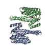
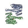
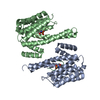
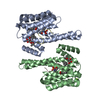
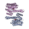
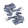
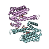
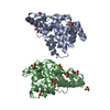
Structure of the LKB1 and 14-3-3 complex
Components
14-3-3 protein zeta/delta
Serine/threonine-protein kinase STK11
Keywords
PROTEIN BINDING/TRANSFERASE / Kinase / Tumor suppressor / Protein Complex / PROTEIN BINDING-TRANSFERASE complex
Function / homology
Function and homology information
positive regulation of vesicle transport along microtubule / intracellular protein-containing complex / Golgi localization / AMPK inhibits chREBP transcriptional activation activity / dendrite extension / synaptic target recognition / activation of protein kinase activity / Golgi reassembly / negative regulation of epithelial cell proliferation involved in prostate gland development / regulation of phosphatidylinositol 3-kinase/protein kinase B signal transduction ...positive regulation of vesicle transport along microtubule / intracellular protein-containing complex / Golgi localization / AMPK inhibits chREBP transcriptional activation activity / dendrite extension / synaptic target recognition / activation of protein kinase activity / Golgi reassembly / negative regulation of epithelial cell proliferation involved in prostate gland development / regulation of phosphatidylinositol 3-kinase/protein kinase B signal transduction / NOTCH4 Activation and Transmission of Signal to the Nucleus / serine/threonine protein kinase complex / epithelial cell proliferation involved in prostate gland development / establishment of Golgi localization / tissue homeostasis / respiratory system process / Energy dependent regulation of mTOR by LKB1-AMPK / tube formation / positive thymic T cell selection / regulation of synapse maturation / Rap1 signalling / vasculature development / regulation of Wnt signaling pathway / anoikis / negative regulation of cold-induced thermogenesis / protein dephosphorylation / positive regulation of axonogenesis / KSRP (KHSRP) binds and destabilizes mRNA / negative regulation of protein localization to nucleus / peptidyl-threonine phosphorylation / cellular response to UV-B / GP1b-IX-V activation signalling / G1 to G0 transition / LRR domain binding / FOXO-mediated transcription of cell death genes / response to ionizing radiation / regulation of dendrite morphogenesis / positive regulation of transforming growth factor beta receptor signaling pathway / establishment of cell polarity / intrinsic apoptotic signaling pathway by p53 class mediator / Regulation of localization of FOXO transcription factors / Interleukin-3, Interleukin-5 and GM-CSF signaling / Activation of BAD and translocation to mitochondria / phosphoserine residue binding / protein kinase activator activity / SARS-CoV-2 targets host intracellular signalling and regulatory pathways / regulation of ERK1 and ERK2 cascade / protein localization to nucleus / cellular response to glucose starvation / SARS-CoV-1 targets host intracellular signalling and regulatory pathways / protein targeting / RHO GTPases activate PKNs / Chk1/Chk2(Cds1) mediated inactivation of Cyclin B:Cdk1 complex / ERK1 and ERK2 cascade / negative regulation of TORC1 signaling / Transcriptional and post-translational regulation of MITF-M expression and activity / axonogenesis / lung development / positive regulation of autophagy / negative regulation of innate immune response / regulation of signal transduction by p53 class mediator / hippocampal mossy fiber to CA3 synapse / TP53 Regulates Metabolic Genes / Translocation of SLC2A4 (GLUT4) to the plasma membrane / protein sequestering activity / Deactivation of the beta-catenin transactivating complex / regulation of protein stability / regulation of cell growth / negative regulation of canonical Wnt signaling pathway / negative regulation of cell growth / Negative regulation of NOTCH4 signaling / positive regulation of protein localization to nucleus / autophagy / p53 binding / melanosome / protein autophosphorylation / intracellular protein localization / T cell receptor signaling pathway / glucose homeostasis / angiogenesis / protein phosphatase binding / blood microparticle / spermatogenesis / DNA-binding transcription factor binding / Regulation of TP53 Activity through Phosphorylation / vesicle / protein phosphorylation / transmembrane transporter binding / non-specific serine/threonine protein kinase / regulation of cell cycle / cilium / cadherin binding / negative regulation of cell population proliferation / protein domain specific binding / protein serine kinase activity / focal adhesion / protein serine/threonine kinase activity / DNA damage response / ubiquitin protein ligase binding / centrosome Similarity search - Function
Serine/Threonine kinase LKB1, catalytic domain / 14-3-3 domain / Delta-Endotoxin; domain 1 / 14-3-3 proteins signature 2. / 14-3-3 protein, conserved site / 14-3-3 proteins signature 1. / 14-3-3 protein / 14-3-3 homologues / 14-3-3 domain / 14-3-3 domain superfamily ...Serine/Threonine kinase LKB1, catalytic domain / 14-3-3 domain / Delta-Endotoxin; domain 1 / 14-3-3 proteins signature 2. / 14-3-3 protein, conserved site / 14-3-3 proteins signature 1. / 14-3-3 protein / 14-3-3 homologues / 14-3-3 domain / 14-3-3 domain superfamily / 14-3-3 protein / Serine/threonine-protein kinase, active site / Serine/Threonine protein kinases active-site signature. / Protein kinase domain / Serine/Threonine protein kinases, catalytic domain / Protein kinase, ATP binding site / Protein kinases ATP-binding region signature. / Protein kinase domain profile. / Protein kinase domain / Protein kinase-like domain superfamily / Up-down Bundle / Mainly Alpha Similarity search - Domain/homology
In the structure databanks used in Yorodumi, some data are registered as the other names, "COVID-19 virus" and "2019-nCoV". Here are the details of the virus and the list of structure data.
Jan 31, 2019. EMDB accession codes are about to change! (news from PDBe EMDB page)
EMDB accession codes are about to change! (news from PDBe EMDB page)
The allocation of 4 digits for EMDB accession codes will soon come to an end. Whilst these codes will remain in use, new EMDB accession codes will include an additional digit and will expand incrementally as the available range of codes is exhausted. The current 4-digit format prefixed with “EMD-” (i.e. EMD-XXXX) will advance to a 5-digit format (i.e. EMD-XXXXX), and so on. It is currently estimated that the 4-digit codes will be depleted around Spring 2019, at which point the 5-digit format will come into force.
The EM Navigator/Yorodumi systems omit the EMD- prefix.
Related info.:Q: What is EMD? / ID/Accession-code notation in Yorodumi/EM Navigator
Yorodumi is a browser for structure data from EMDB, PDB, SASBDB, etc.
This page is also the successor to EM Navigator detail page, and also detail information page/front-end page for Omokage search.
The word "yorodu" (or yorozu) is an old Japanese word meaning "ten thousand". "mi" (miru) is to see.
Related info.:EMDB / PDB / SASBDB / Comparison of 3 databanks / Yorodumi Search / Aug 31, 2016. New EM Navigator & Yorodumi / Yorodumi Papers / Jmol/JSmol / Function and homology information / Changes in new EM Navigator and Yorodumi
 Movie
Movie Controller


 Open data
 Basic information
 Components
 Keywords
 Function and homology information
 Homo sapiens (human)
 X-RAY DIFFRACTION /
  Authors
 Citation
 Structure visualization
 Downloads & links
 Other downloads
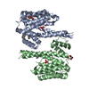


 PDBj
PDBj


 Assembly
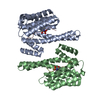

 Mass: 18.015 Da / Num. of mol.: 18 / Source method: isolated from a natural source / Formula: H2O
 Sample preparation
 / Beamline: BL17U / Wavelength: 0.97915 Å
 Processing